Author: Dr. Prince

  • 3 Best Nursing Care Plans for Pressure Ulcers Based on Diagnosis

    This article discusses Nursing Care Plans for Pressure Ulcers plus its causes, symptoms, preventions, treatments, and interventions.

    Permalink: https://customnursingassignments.com/nursing-care-plans-for-pressure-ulcers

    customnursingassignments.com has the top and most qualified writers to help with any of your assignments. All you need to do is place an order with us.(Nursing Care Plans for Pressure Ulcers)

    Disclaimer: The information presented in this article is not medical advice; it is meant to act as a quick guide to nursing students for learning purposes only and should not be applied without an approved physician’s consent. Please consult a registered doctor in case you’re looking for medical advice.

    Introduction

    Pressure ulcers (also known as pressure sores or bedsores) are injuries to the skin and underlying tissue, primarily caused by prolonged pressure on the skin. Ulcers are more common on bony prominences with less cushioning between bone and skin (such as elbows, coccyx, and heels). They can also manifest themselves in other parts of the body. They can happen to anyone but usually affect people confined.

    These breaks may not heal on their own because bacteria on the person’s skin or clothing colonize deep into the wet wound bed, causing it to become infected. The assessment and diagnosis of pressure ulcers, as well as risk factors, consequences, and therapies, are discussed in this blog post.

    Pressure ulcers can develop over hours or days. Most sores heal with treatment, but some never heal completely. You can take steps to help prevent bedsores and help them heal.

    Pressure Ulcers

    A pressure ulcer, also known as bedsore or decubitus ulcer, is a localized skin injury caused by tissue compression between bony prominences and hard surfaces like a mattress. Pressure, in combination with friction, shearing forces, and moisture, causes them. Small blood arteries are compressed by the pressure, resulting in poor tissue perfusion. Reduced blood flow induces tissue hypoxia, which results in cellular death.

    Common sites of pressure ulcers

    Pressure ulcers on the skin across the following locations are common in wheelchair users:

    • Buttocks or tailbone
    • Spine and shoulder blades
    • Backs of arms and legs where they rest against the chair
    • Backs of arms and legs where they rest against the chair
    • Pressure ulcers can occur in people who have to stay in bed for long periods of time.
    • The back or sides of the head.
    • The shoulder blades.
    • The hip, lower back, or tailbone.
    • The skin behind the knees, the heels, and the ankles.

    Types of Pressure Ulcers

    Bedsores, decubitus ulcers, and trochanteric bursitis are the most prevalent types of pressure ulcers. They don’t all cause deep tissue injury. They can be further divided into the following categories:

    Dry ulcers

    When scraped, the skin is dry and does not bleed. The tissue’s blood supply may be harmed or destroyed. The area feels firm to the touch.

    Wet/Moist ulcers

    A damp or moist feeling is common with this form of pressure ulcer. When the wounded region is scraped, the skin may bleed, and inflammation manifests itself as redness, swelling, and warmth.

    Cold ulcers

    When this sort of ulcer is touched, it does not cause pain or bleeding. Instead, due to a lack of blood flow, the tissue will be cold (also known as poor perfusion). It’s possible that your skin will appear pale and even frosty.

    Sterile ulcers

    An ulcer of this sort is red, swollen, and heated to the touch. The tissue’s blood supply has been cut off or diminished, but there are no microorganisms in the area.

    Will Not Heal Pressure ulcers

    This type of pressure sore has not responded to treatment and will not heal without surgical intervention.

    Symptoms of pressure ulcers

    Pressure ulcers can affect any part of the body that’s put under pressure. They’re most common on bony parts of the body, such as the heels, elbows, hips and base of the spine.

    They often develop gradually but can sometimes form in a few hours.

    Early symptoms

    • Part of the skin becoming discoloured – people with pale skin tend to get red patches, while people with dark skin tend to get purple or blue patches.
    • Discoloured patches not turning white when pressed
    • A patch of skin that feels warm, spongy or hard
    • Pain or itchiness in the affected area

    Later symptoms

    • An open wound or blister – a category two pressure ulcer
    • A deep wound that reaches the deeper layers of the skin – a category three pressure ulcer
    • A very deep wound that may reach the muscle and bone – a category four pressure ulcer

    Risk factors of pressure ulcers

    Suppose you have trouble moving and changing positions when seated or in bed; you’re more likely to acquire bedsores. The following are some of the risk factors:

    Immobility. This could be due to a variety of factors, including poor health, spinal cord injury, and others.

    Incontinence. When skin is exposed to urine and stool for an extended period of time, it becomes more susceptible.

    Lack of sensory perception. Sensation loss can occur as a result of spinal cord injury, neurological illnesses, and other conditions. Lack of awareness of warning indicators and the need to shift positions can stem from an inability to feel pain or discomfort.

    Medical conditions affecting blood flow. Diabetes and vascular illness, both of which can impair blood flow, might increase the risk of tissue damage such as bedsores.

    Deficiency in nutrients and hydration. To maintain healthy skin and avoid tissue breakdown, people require enough water, calories, protein, vitamins, and minerals in their daily diets.

    Stages of Pressure Ulcers

    The stages are determined by the depth of the sores, which has an impact on how they are treated.

    If caught early, these sores are likely to heal in a few days with little fuss or pain. Without treatment, they can get worse.

    When the sore shrinks and pink tissue appears along the sides, you’ll know they’re getting better.

    The stages include:

    Stage 1
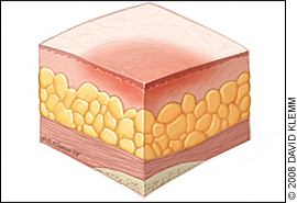
    This is the mildest stage. These pressure sores only affect the upper layer of your skin.

    Symptoms: Pain, burning, or itching are common symptoms. The spot may also feel different from the surrounding skin: firmer or softer, warmer or cooler.

    You may notice a red area on your skin. If you have darker skin, the discoloured area may be harder to see. The spot doesn’t get lighter when you press on it, or even 10 to 30 minutes after you stop pressing. This means less blood is getting to the area.

    What to do: The first and most important thing to do with any pressure sore is to stop the pressure. Change your position or use foam pads, pillows, or mattresses.

    If you spend a lot of time in bed, try to move at least once every 2 hours. If you’re sitting, move every 15 minutes. You may need someone to help you.

    Wash the sore with mild soap and water and dry it gently.

    It may help to eat a diet high in protein, vitamins A and C, and the minerals iron and zinc. These are all good for your skin. Also, drink plenty of water.

    Recovery time: A Stage 1 pressure sore may go away in as little as 2 or 3 days.

    Stage 2
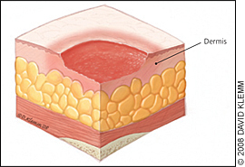
    This happens when the sore digs deeper below the surface of your skin.

    Symptoms: Your skin is broken, leaves an open wound, or looks like a pus-filled blister.

    The area is swollen, warm, and/or red. The sore may ooze clear fluid or pus. And it’s painful.

    What to do: Follow the same steps for Stage 1. You should also clean the wound with water or a salt-water solution and dry it gently. This may hurt, so ask your doctor if you should take a pain reliever 30 to 60 minutes before cleaning.

    Keep the sore covered with a see-through dressing or moist gauze. If you see signs of an infection (such as pus, fever, or redness), tell your doctor.

    Recovery time: A Stage 2 pressure sore should get better in 3 days to 3 weeks.

    Stage 3
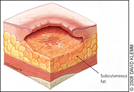
    These sores have gone through the second layer of skin into the fat tissue.

    Symptoms: The sore looks like a crater and may have a bad odour. It may show signs of infection: red edges, pus, odour, heat, and/or drainage. The tissue in or around the sore is black if it has died.

    What to do: Stage 3 sores will need more care. Talk to your doctor. They may remove any dead tissue and prescribe antibiotics to fight infection. You may also be able to get a special bed or mattress through your insurance.

    Recovery time: A Stage 3 pressure sore will take at least one month and up to 4 months to heal.

    Stage 4
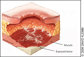
    These sores are the most serious. Some may even affect your muscles and ligaments.

    Symptoms: The sore is deep and big. The skin has turned black and shows signs of infection — red edges, pus, odour, heat, and/or drainage. You may be able to see tendons, muscles, and bones.

    What to do: Tell your doctor right away. These wounds need immediate attention, and you may need surgery.

    Recovery time: A Stage 4 pressure sore could take anywhere from 3 months or much longer, even years, to heal.

    Causes of Pressure Ulcers

    Pressure ulcers are caused by pressure against the skin that limits blood flow to the skin. Limited movement can make skin vulnerable to damage and lead to the development of pressure ulcers.

    Three primary contributing factors for pressure ulcers are:

    Pressure – Constant pressure on any part of your body can lessen the blood flow to tissues. Blood flow is essential for delivering oxygen and other nutrients to tissues. Without these essential nutrients, skin and nearby tissues are damaged and might eventually die. For people with limited mobility, this kind of pressure tends to happen in areas that aren’t well-padded with muscle or fat and that lie over a bone, such as the spine, tailbone, shoulder blades, hips, heels and elbows.

    Friction – Friction occurs when the skin rubs against clothing or bedding. It can make fragile skin more vulnerable to injury, especially if the skin is also moist.

    Shear – Shear occurs when two surfaces move in the opposite direction. For example, when a bed is elevated at the head, you can slide down in bed. As the tailbone moves down, the skin over the bone might stay in place — essentially pulling in the opposite direction.

    Complications

    The following are some of the life-threatening complications of pressure ulcers:

    Cellulitis – Cellulitis is an infection of the skin and soft tissues that surround it. It might cause the affected area to become heated, red, and swollen. People with nerve damage frequently do not experience pain in the cellulitis-affected area.

    Bone and joint infections – A pressure sore infection can spread to joints and bones. Infections in the joints (septic arthritis) can cause cartilage and tissue damage. Joint and limb function can be harmed by bone infections (osteomyelitis).

    Cancer – Squamous cell carcinoma can arise from long-term, non-healing wounds (Marjolin’s ulcers).

    Sepsis – A skin ulcer can cause sepsis in rare cases.

    Preventions for pressure ulcers

    It can be difficult to completely prevent pressure ulcers, but there are some things you or your care team can do to reduce the risk.

    These include:

    1. Regularly changing your position – if you’re unable to change position yourself, a relative or carer will need to help you
    2. Checking your skin every day for early signs and symptoms of pressure ulcers – this will be done by your care team if you’re in a hospital or care home.
    3. Having a healthy, balanced diet that contains enough protein and a good variety of vitamins and minerals.
    4. Stopping smoking – smoking makes you more likely to get pressure ulcers because of the damage caused to blood circulation.

    As you continue, customnursingassignments.com has the top and most qualified writers to help with any of your assignments. All you need to do is place an order with us. (Nursing Care Plans for Pressure Ulcers)

    Nursing Care Plans for Pressure Ulcers
    Nursing Care Plans for Pressure Ulcers

    Nursing Diagnosis for Pressure Ulcers

    Diagnosis involves;

    1. Physical examination to check for signs such as erythema and fluid seeping from the lesion.
    2. Impaired circulation—what could be the cause? Is it a vascular problem or a result of your position?
    3. To look for signs of infection, such as redness, swelling, discomfort, and a raised temperature.
    4. Lab tests to see if the patient has an infection or another ailment. Urine analysis, cultures, and blood counts are examples of these assays.

    Nursing Care Plans for Pressure Ulcers

    Pressure ulcers stage I through III can be managed with aggressive local wound treatment and proper nutritional support, while stage IV pressure ulcers usually require surgical intervention.

    Measuring the contributing variables leading to a lack of tissue perfusion, assessing the extent of the injury, increasing medication compliance, and preventing additional harm is all part of nursing care for clients with pressure ulcers (bedsores).

    Nursing Care Plan Based on Impaired Skin Integrity Diagnosis – Nursing Care Plans for Pressure Ulcers

    Nursing Diagnosis: Impaired Skin Integrity

    It may be related to:

    • Chronic disease state.
    • Extreme of ages.
    • Imbalanced nutritional state.
    • Impaired cognition.
    • Impaired sensation.
    • Immobility.
    • Immunological deficit.
    • Incontinence.
    • Mechanical factors (friction, pressure, shear).
    • Moisture.
    • Poor circulation.
    • Pronounced body prominence.
    • Radiation.

    Evidence of on Impaired Skin Integrity Diagnosis

    • Destruction of skin layers.
    • Disruption of skin surfaces.
    • Drainage of pus.
    • Invasion of body structures.
    • Pressure ulcer stages:
      • Deep tissue injury (new stage):

    Purple or localized maroon area of intact skin or blood-filled blister resulting from pressure damage of underlying soft tissue.

    • Stage I:

    The epidermis is intact.

    Non-branch able erythema of intact skin. Discolouration of the skin, warmth, edema, induration or hardness may also be used as indicators, particularly on individuals with darker skin.

    • Stage II:

    Partial-thickness skin loss involving epidermis, dermis, or both. The ulcer is superficial and presents clinically as an abrasion or blister.

    • Stage III:

    Full-thickness skin loss involving damage to or necrosis of subcutaneous tissue that may extend down to, but not through, underlying fascia.

    Slough may be present; it may include undermining and tunnelling.

    As you continue, customnursingassignments.com has the top and most qualified writers to help with any of your assignments. All you need to do is place an order with us. (Nursing Care Plans for Pressure Ulcers)

    Nursing Care Plans for Pressure Ulcers
    Nursing Care Plans for Pressure Ulcers
    • Stage IV:

    Extensive destruction, tissue necrosis or damage to muscle, bone or supporting structures, with or without full-thickness skin loss.

    Undermining and tunnelling may develop.

    • Unstageable:

    Full-thickness tissue loss in which actual depth of ulcer is completely obstructed by slough or eschar in the wound bed.

    Desired Outcomes

    The client will get stage-appropriate wound care and has controlled risk factors for the prevention of additional ulcers.

    The client will experience healing of pressure ulcers and experiences pressure reduction.

    Nursing InterventionsRationale
    Assess the specific risk factors for pressure ulcers:Even clients with an existing pressure ulcer continue to be at risk for further injury. Nurses should consider all potential risk factors for pressure ulcers development.
    Determine the client’s age and general condition of the skin.Elderly clients have less elastic skin, less moisture, less padding and have thinning of the epidermis, making it more prone to skin impairment.
    Assess the client’s nutritional status, including weight, weight loss, and serum albumin levels, if indicated.A severe protein depletion has an albumin level of less than 2.5 g/dL. Clients with pressure ulcers lose large amounts of protein in wound exudates and may require 4000 kcal/day or more to remain anabolic.
    Assess for a history of preexisting chronic diseases (e.g., diabetes mellitus, acquired immune deficiency syndrome, Guillain-barré syndrome, peripheral and/or cardiovascular disease).Clients with chronic diseases typically exhibit multiple risk factors that predispose them to pressure ulceration. These include poor nutrition, poor hydration, incontinence, and immobility.
    Assess the skin on admission and daily for an increasing number of risk factors.The incidence of skin breakdown is directly related to the number of risk factors present.
    Assess for a history of radiation therapy.Irradiated skin becomes thin and brittle, may have less blood supply, and is at a higher risk for skin breakdown.
    Assess the client’s awareness of the sensation of pressure. Usually, people shift their weight off pressure areas every few minutes; this occurs more or less automatically, even during sleep. Clients with decreased sensation are unaware of unpleasant stimuli and do not shift weight, thereby exposing the skin to excessive pressure.
    Assess for fecal and urinary incontinence. The urea in urine turns into ammonia within minutes and is erosive to the skin. At the same time, the stool may contain enzymes that cause skin breakdown. Diapers and incontinence pads with plastic liners trap moisture and speed up breakdown.
    Assess client’s ability to move (shift weight while sitting, turn over in bed, move from the bed to a chair). Immobility is a huge risk factor for pressure ulcer development among adult hospitalized clients.
    Assess for environmental moisture (excessive perspiration, high humidity, wound drainage). Moisture may contribute to skin maceration.
    Assess the amount of shear (pressure exerted laterally) and friction (rubbing) on the client’s skin. Shearing forces are most commonly noted on the sacrum, scapulae, heels, and elbows from skin-sheet friction, from semi-Fowler’s position and repositioning, and from lift sheets.
    Assess the surface that the clients spend a majority of time on (mattress for bedridden clients, cushion for clients in wheelchairs). Clients who spend the majority of their time on one surface need a pressure reduction or pressure relief device to reduce the risk of skin breakdown.
    Assess the skin over bony prominences (sacrum, trochanters, scapulae, elbows, heels, inner and outer malleolus, inner and outer knees, back of the head).These areas are at the highest risk for breakdown resulting from tissue ischemia from compression against a hard surface.
    Use an objective tool for pressure ulcer risk assessment: Braden scale. Norton scale.The Braden scale is the most widely used risk assessment. It consists of six subscales, namely: activity, mobility, moisture, nutrition, sensory perception, and friction. Acute care: Assessment should be carried out on all patients on admission and every 24 to 48 hours or sooner if the patient’s condition changes. Long-term care: Assess on admission, weekly for four weeks, then quarterly and whenever the resident’s condition changes.
    Assess the client’s level of pain, especially related to dressing change and procedures.Prophylactic pain medication may be indicated.
    Assess and stage the pressure ulcers.Staging is essential because it determines the treatment plan. Staging should be assessed at each dressing stage. It reflects whether the epidermis, dermis, fat, muscle, bone, or joint is exposed. If the ulcer is covered with necrotic tissue (eschar), it cannot be accurately staged. Stage I ulcers are difficult to detect in darkly pigmented skin. The use of mirrors or a penlight may be helpful.
    Determine the condition of the wound or wound bed. 
    Presence of necrotic tissue.Necrotic tissue is a tissue that is dead and eventually must be removed before healing can take place. Necrotic tissue exhibits a wide range of appearances: black, brown, leathery, hard, shiny, thin, tough, white.
    Colour.The colour of tissue is an indication of tissue viability and oxygenation. White, gray, or yellow eschar may be present in stage II and III ulcers. Eschar may be black in stage IV ulcers.
    Odour.An odour may arise from infection present in the wound; it may also arise from the necrotic tissue. Some local wound care products may create or intensify the odours and should be distinguished from wound or exudate odours.
    Viability of bone, joints, or muscle.In stage IV pressure ulcers, these may be apparent at the base of the ulcer. Wounds may demonstrate multiple stages or characteristics in a single wound.
    Measure the size of the ulcer, and note the presence of undermining.The ulcer dimensions include length, width, and depth. An ulcer begins in the deepest tissue layers before the skin breaks down. Hence the opening of the skin’s surface may not represent the true size of the ulcer.
    Assess the condition of wound edges and surrounding tissue.The surrounding tissue may be healthy or may have various degrees of impairment. Healthy tissue is necessary for the use of local wound care products requiring adhesion to the skin. The presence of healthy tissue demarcates the boundaries of the pressure ulcer.
    Assess the wound exudate.Exudate is a normal part of wound physiology and must be differentiated from pus which is an indication of infection. Exudate may contain serum, blood, and white blood cells and may appear clear, cloudy, or blood-tinged. The amount may vary from a few cubic centimetres, which are easily managed with dressings, to copious amounts not easily managed. Drainage is considered excessive when dressing changes are needed more often than every 6 hours.
    Assess ulcer healing using a pressure ulcer scale for healing (PUSH) tool.This tool provides standardization in the measurement of wound healing. It quantifies surface area, exudate, and the type of wound tissue.
    Provide local wound care:
    Stage I:
    Apply a topical vasodilator (e.g., Proderm)It increases skin circulation.
    Apply a flexible hydrocolloid dressing (e.g., Duoderm) or a vapour-permeable membrane dressing (Tegaderm).It prevents shear and friction.
    Apply a vitamin-enriched emollient to the skin every shift.It moisturizes the skin.
    Stage II:
    Apply Alginates (Sorbsan, Kalginate, Kaltostat).Alginate dressings are a type that is highly absorbent and so can absorb the fluid (exudate) that is produced by some ulcers. These are often used for ulcers with moderate-to-heavy exudate.
    Apply hydrocolloids or a vapour-permeable membrane dressing.Hydrocolloids are used to promote healing and wound debridement. They are not advised to use for heavy-exudate-producing wounds.
    Apply gauze with sodium chloride solution.This maintains a moist environment but requires multiple dressing changes. Dressings must be removed while still wet. Dressings absorb small amounts of drainage.
    Apply Hydrogels (Carrasyn V, Aqua Skin).Hydrogels provide moisture to dry, sloughy or necrotic wounds and assists autolytic debridement. It can be used on wounds with low exudate and is usually used for shallow ulcers without exudates.
    Stage III and IV:
    Foams.Different foams have different levels of absorbency. They are best used on granulating wounds. Foams lessen odour and repel bacteria and water.
    Gauze with sodium chloride solution.This maintains a moist environment but requires multiple dressing changes as described for stage II.
    Wound fillers.Wound fillers are used as a primary dressing and to pack wounds, maintain a moist environment.
    Autolytic debridement.Using a hydrocolloid or hydrogel, these create a moist wound interface that enhances the activity of endogenous proteolytic enzymes within the wound, liquefying and separating necrotic tissue from healthy tissue.
    Sharp or surgical debridement.This procedure removes the necrotic tissue and senescent cells that slow down the tissue repair process, converting a chronic wound into an acute one in the process.
    Mechanical debridement.It involves allowing a traditional gauze-type dressing to dry out and adhere to the surface of the wound before manually removing the dressing, debriding any tissue attached to it.
    Electrical stimulation.Stimulation of many cellular processes improves healing.
    Biosurgery.Therapeutic use of live blowfly larvae (maggots) for quick debridement.
    Topical growth factors.Nerve-growth factors, colony-stimulating factors, and fibroblast growth factors are found to be effective in treating diabetic and venous ulcers.
    Negative pressure wound therapy.A wound dressing system that continuously or intermittently apply a subatmospheric pressure to the surface of a wound to assist healing.
    Enzymatic debridement (chlorophyll, collagenase, papain).Enzymatic debridement uses proteolytic enzymes to remove necrotic tissue. These agents work by selectively digesting the collagen portion of the necrotic tissue. Care should be taken to prevent damage to surrounding healthy tissues.

    Nursing Care Plan Based on Risk for Infection Diagnosis – Nursing Care Plans for Pressure Ulcers

    Nursing Diagnosis: Risk for Infection

    Risk factors

    Poor nutritional status.

    The proximity of sacral wounds to the perineum.

    Open pressure ulcer.

    Desired Outcomes

    • The client will maintain a normal body temperature.
    • The client will remain free of local or systemic infections, as evidenced by the absence of copious, foul-smelling wound exudate.
    Nursing InterventionsRationale
    Assess the client’s nutritional status.Clients who seriously lack nutrition (serum albumin <2.5 mg/dl) are at risk of developing infection produced by a pressure ulcer. Also, clients with pressure ulcers lose tremendous amounts of protein in wound exudate and may require 4000 kcal/day or more to remain anabolic.
    Assess the client for unexplained sepsis.When septic workup is done, the pressure ulcer must be considered a possible cause.
    Assess for urinary and fecal incontinence.Sacral wounds, because of their proximity to the perineum, are at the highest risk for infection caused by urine or fecal contamination. It is sometimes difficult to isolate the wound from the perineal area.
    Assess pressure ulcers for odour, the colour of tissue, and drainage.Foul-smelling pressure ulcer may indicate an infection; Infected tissue usually has a gray-yellow appearance without evidence of pink granulation tissue; The presence of exudate that is clear to straw-coloured is normal. While purulent green or yellow drainage in large amounts indicates an infection.
    Assess the client’s temperature.Fever is considered a temperature above 100.4 degrees F (38 degrees C) indicate a presence of infection unless the client is immunocompromised or diabetic.
    Monitor the client’s white blood cell count (WBC).Elevated WBC counts indicate an infection, although, in very old individuals, the WBC count may rise only slightly during infection, indicating a diminished marrow reserve.
    Obtain wound cultures, if indicated.All pressure ulcers are colonized because skin normally has flora that will be found in an open skin lesion; however, all pressure ulcers are not infected. Infection is present when there is copious, foul-smelling, purulent drainage and the client has other signs of infection (fever, increased pain) and bacteria count greater than 105. Swab cultures are not recommended. Rather, tissue biopsy should be used to quantify and qualify the aerobic and anaerobic organisms present.
    Consult with a dietitian for assistance with a high-protein, high-calorie diet.A High-calorie, high-protein diet may be recommended to help in healing and resist infection.
    Provide thorough perineal hygiene after each episode of incontinence.This can lessen pathogens in the area of sacral pressure ulcers.
    Provide hydrotherapy, if indicated.It is used to achieve wound cleansing and to promote good circulation.
    Administer antibiotics as prescribed.Complicated wounds may develop cellulitis or sepsis, requiring antibiotic therapy. Oral antibiotics or topical silver sulfadiazine can be effective.
    Provide local wound care as prescribed.The type and level of wound treatment depend on the staging of the ulcer and the type of infection present.

    As you continue, customnursingassignments.com has the top and most qualified writers to help with any of your assignments. All you need to do is place an order with us. (Nursing Care Plans for Pressure Ulcers)

    Nursing Care Plans for Pressure Ulcers
    Nursing Care Plans for Pressure Ulcers

    Nursing Care Plan Based on Risk For Ineffective Health Maintenance Diagnosis – Nursing Care Plans for Pressure Ulcers

    Nursing Diagnosis: Risk for Ineffective Health Maintenance

    Risk factors

    Impaired functional status.

    Lack of previous similar experience.

    Need for long-term pressure management.

    Possible need for special equipment.

    Desired Outcomes

    Client and caregiver will verbalize understanding of the following aspects of home care: nutrition, pressure relief, wound care, and incontinence management.

    Client and caregiver will verbalize ability to cope adequately with an existing situation, provide support/monitoring as indicated.

    Nursing InterventionsRationale
    Assess the client’s and caregiver’s knowledge of and ability to provide local wound care.Client’s are no longer kept hospitalized until pressure ulcers have healed. The need for local wound care may continue at home for weeks to months.
    Assess the client’s and caregiver’s understanding of the prevention of further pressure ulcer development.Clients who are immobile will need frequent repositioning to lessen the risk fro breakdown in those areas that are intact.
    Assess the client’s and caregiver’s understanding of the long-term nature of wound healingPressure ulcers may take weeks to months to heal, even under ideal circumstances. Wound heal from the base of the ulcer up and from the edges of the ulcer toward the center. Palliative wound care may be appropriate for a clean, chronic, non-healing wound.
    Assess the client’s and caregiver’s understanding of the relationship between incontinence and further skin breakdown or complication of healing.Managing incontinence may be the most difficult aspect of home management and is often the reason for nursing home placement are made.
    Assess the client’s and caregiver’s understanding of and ability to provide a High-calorie, high-protein diet throughout the course of wound healing.Clients may require enteral feeding (through gastronomy tube, nasogastric tube feedings, or the oral route), which requires knowledge of preparation and the use of special equipment.
    Assess for the availability of a pressure reduction or pressure-relief surface.A client may take a thick, dense foam mattress home from the hospital to place it on their own bed. Rental provisions of low-air-loss beds (e.g., KinAir, Flexicare) and air-fluidized therapy beds (FluidAir, Skytron, Clintron) may be arranged but often pose a financial difficulty because few payer sources will cover the cost of these beds in the home.
    Educate the client and the caregiver to report the following signs indicating wound infection: Fever, malaise, chills, foul-smelling odour, purulent drainage.Early detection prompts immediate intervention.
    Educate the client and the caregiver in managing incontinence (e.g., use of moisture barrier ointments, use of underpads, use of external catheters).Teaching proper techniques can prevent leakage and skin problems. Reusable products such as underpads or linen protectors made of cloth with a waterproof lining are better for the client’s skin and are more economical but require laundering. Moisture barrier ointments protect intact skin from excoriation.
    Educate the client and the caregiver regarding local wound care, and allow for a return demonstration.This will allow the client to use new information immediately, thus enhancing retention. Immediate feedback allows the learner to make corrections rather than practice the skill incorrectly.
    Provide written instructions with listed resources.Long-term management requires specific written plans to enhance adherence o treatment. Several internet resources provide lay education.
    Involve a social worker or case manager.Referral helps the client and family determine whether placement in an extended care facility is needed. Because many clients with pressures are older, it is often an older spouse who is available to provide care; as a result of the intensive nursing care needs of these clients, discharge to home is often unrealistic.
    Consult a wound specialist to evaluate care in the home.Besides evaluating the ability to deliver care, the specialist may be useful in securing specialty treatment.
    Educate the client and the caregiver on the importance of pressure reduction and relief (e.g., turning schedule, use of specialty beds, use of relief surface where the client sits).Information can nurture enhanced adherence to pressure ulcer treatment guidelines.
    Discuss with the client and caregiver the possible need for respite care.Long-term responsibility for client care in the home is burdening; those providing care may need help to understand that their needs for relaxation are important to the maintenance of health and should not be viewed as avoidance of the responsibility.
    Discuss with the client and caregiver the need for in-home nursing care or homemaker services.These provide all or part of the client’s care and can be economical to the client. Also, keeping the client in his or her own environment lessen the risk for hospital-acquired infection and keeps the client in familiar surrounding.

    Summary

    Patients of all ages are susceptible to pressure ulcers, which are particularly common among diabetics. Prolonged bed rest (at least 8 hours at a time), starvation, dehydration, poor circulation, edema, and lack of mobility are the major causes. Pressure ulcer severity is divided into four stages:  stage I, stage II, stage III and stage IV.

    Proper wound care products, such as topical negative pressure dressings, support surfaces, padding, and bandages, are the greatest strategy to avoid ulcer formation. Giving patients well-balanced food, as well as keeping them hydrated and mobile, will aid in their recovery.

    Related FAQs – Nursing Care Plans for Pressure Ulcers

    1. What are the 4 stages of pressure ulcers?

    These are:

    • Stage 1. The area looks red and feels warm to the touch. …
    • Stage 2. The area looks more damaged and may have an open sore, scrape, or blister. …
    • Stage 3. The area has a crater-like appearance due to damage below the skin’s surface.
    • Stage 4. The area is severely damaged and a large wound is present.

    2. What is the best treatment of pressure ulcers?

    Clean open sores with water or a saltwater (saline) solution each time the dressing is changed. Putting on a bandage. A bandage speeds healing by keeping the wound moist. It also creates a barrier against infection and keeps skin around it dry.

    3. What are the common symptoms of a pressure ulcer?

    Warning Signs and Symptoms of Pressure Ulcers

    • A change in the colour of the skin. …
    • Reddening of the skin that does NOT turn white when you press it. …
    • Any unusual changes in skin texture may be related to pressure damage. …
    • A patch of skin that feels cooler or warmer to the touch than others.
    • A sore or itchy patch of skin.

    4. What antibiotics are used for pressure ulcers?

    Amoxicillin-potassium clavulanate is a naturally occurring beta-lactam structurally similar to the penicillin nucleus. This antibiotic group of beta-lactam/beta-lactamase combination has demonstrated a broad-spectrum activity; therefore, it is frequently used for the treatment of infected pressure ulcers.

    Read More:

    https://customnursingassignments.com/nursing-care-plan-for-syncope/
    https://customnursingassignments.com/nursing-care-plans-for-traumatic-brain-injuries/
  • Nursing Care Plan For Syncope – Best Nursing Care Plans(2022)

    This article discusses Nursing Care Plan For Syncope plus its causes, symptoms, preventions, treatments, and interventions.

    Permalink: https://customnursingassignments.com/nursing-care-plan-for-syncope

    customnursingassignments.com has the top and most qualified writers to help with any of your assignments. All you need to do is place an order with us.(Nursing Care Plan For Syncope)

    Disclaimer: The information presented in this article is not medical advice; it is meant to act as a quick guide to nursing students for learning purposes only and should not be applied without an approved physician’s consent. Please consult a registered doctor in case you’re looking for medical advice.

    Introduction

    Syncope is the medical word for passing out or fainting. It’s caused by a temporary decrease in blood supply to the brain.

    Syncope can occur if your blood pressure drops suddenly, your heart rate drops, or the amount of blood in different parts of your body fluctuates. If you pass out, you’ll most likely wake up cognizant and alert soon after, but you might be disoriented for a while.

    It is characterized by an abrupt loss of awareness and muscular tone caused by a transient lack of blood flow to the brain. Low blood pressure, dehydration, or rising too rapidly from a sitting or lying down position can contribute to the illness.

    Disclaimer: The information presented in this article is not medical advice; it is meant to act as a quick guide to nursing students for learning purposes only and should not be applied without an approved physician’s consent. Please consult a registered doctor in case you’re looking for medical advice.

    Types of Syncope

    Syncope can take several forms. What type a person has is determined by the cause of the issue.

    Vasovagal Syncope (Cardio-neurogenic Syncope)

    Vasovagal syncope is the most common type of syncope. It is caused by a sudden drop in blood pressure, which causes a decrease in blood flow to the brain. When you stand up, gravity causes blood to settle in the lower part of your body, below your diaphragm. When that happens, the heart and autonomic nervous system (ANS) work to keep your blood pressure stable.

    Some patients with vasovagal syncope have a condition called orthostatic hypotension. This condition keeps the blood vessels from getting smaller (as they should) when the patient stands. This causes blood to collect in the legs and leads to a quick drop in blood pressure.

    Situational syncope

    Situational syncope is a type of vasovagal syncope. It happens only during certain situations that affect the nervous system and lead to syncope. Some of these situations are:

    1. Dehydration
    2. Intense emotional stress
    3. Anxiety
    4. Fear
    5. Pain
    6. Hunger
    7. Use of alcohol or drugs
    8. Hyperventilation (breathing in too much oxygen and getting rid of too much carbon dioxide too quickly)
    9. Coughing forcefully, turning the neck, or wearing a tight collar (carotid sinus hypersensitivity)

    Postural syncope (Postural Hypotension)

    Postural syncope is caused by a sudden drop in blood pressure due to a quick change in position, such as lying down to standing. Certain medications and dehydration can lead to this condition. Patients with this type of syncope usually have changes in their blood pressure that cause it to drop by at least 20 mmHg (systolic/top number) and at least ten mmHg (diastolic/bottom number) when they stand.

    Cardiac syncope is caused by a heart or blood vessel condition that affects blood flow to the brain. These conditions can include an abnormal heart rhythm (arrhythmia), obstructed blood flow in the heart due to structural heart disease (the way the heart is formed), blockage in the cardiac blood vessels (myocardial ischemia), valve disease, aortic stenosis, blood clot, or heart failure. If you have cardiac syncope, it is essential to see a cardiologist for proper treatment.

    Neurologic Syncope

    Neurologic syncope is caused by a neurological condition such as seizure, stroke, or transient ischemic attack (TIA). Other less common conditions that lead to neurologic syncope include migraines and normal pressure hydrocephalus.

    Postural Orthostatic Tachycardia Syndrome (POTS)

    Postural-Orthostatic Tachycardia Syndrome is caused by a fast heart rate (tachycardia) that happens when a person stands after sitting or lying down. The heart rate can speed up by 30 beats per minute or more. The increase usually happens within 10 minutes of standing. The condition is most common in women, but it can also occur in men.

    Symptoms of Syncope

    The most common symptoms of syncope include:

    • Blacking out
    • Feeling lightheaded
    • Falling for no reason
    • Feeling dizzy
    • Feeling tired or dizzy
    • Fainting, especially after eating or exercising
    • Feeling unsteady or weak when standing
    • Changes in vision, such as seeing spots or having tunnel vision
    • Headaches

    Many times, patients feel an episode of syncope coming on. They have what are called “premonitory symptoms,” such as feeling lightheaded, nauseous, and heart palpitations (irregular heartbeats that feel like “fluttering” in the chest). If you have syncope, you will likely be able to keep from fainting if you sit or lie down and put your legs up if you feel these symptoms.

    Syncope can be a sign of a more severe condition. So, it is essential to get treatment right away after you have an episode of syncope. Most patients can prevent problems with syncope once they get an accurate diagnosis and proper treatment.

    Causes Syncope

    Syncope is a symptom that various illnesses can cause, ranging from benign to life-threatening. Syncope can be caused by a variety of non-life-threatening reasons such as overheating, dehydration, intense sweating, tiredness, or blood pooling in the legs owing to fast changes in body position. It’s critical to figure out what’s causing your syncope and whether you have any underlying issues.

    Syncope can also be caused by significant heart disorders such as bradycardia, tachycardia, or blood flow restriction.

    How is syncope diagnosed?

    If you have syncope, you should see your doctor, who can refer you to a syncope specialist for a complete evaluation.

    The evaluation begins with a careful review of your medical history and a physical exam. Your doctor will ask you detailed questions about your symptoms and syncope episodes, including whether you have any symptoms before you faint and when and where the attacks happen.

    You may then have one or more tests to help your doctor determine the cause of your syncope. These tests check things like the condition of your heart, how fast your heart is beating (heart rate), the amount of blood in your body (blood volume), and blood flow in different positions.

    Tests to determine causes of syncope include:

    Laboratory testing: Blood work to check for anemia or metabolic changes.

    Electrocardiogram (EKG or ECG): A test that records the electrical activity of your heart. Electrodes (small sticky patches) are applied to your skin to collect this information.

    Exercise stress test: A test that uses an ECG to record your heart’s electrical activity while you are active. This is done on a treadmill or stationary bike, which helps you reach a target heart rate.

    Ambulatory monitor: You will wear a monitor that uses electrodes to record information about your heart’s rate and rhythm.

    Echocardiogram: A test that uses high-frequency sound waves to create an image of the heart structures.

    Tilt table (head-up tilt test): A test that records your blood pressure and heart rate on a minute-by-minute or beat-by-beat basis while the table is tilted to different levels as you stay head-up. The test can show abnormal cardiovascular reflexes that cause syncope.

    Blood volume determination: A test to see if you have the right amount of blood in your body, based on your gender, height, and weight. A small amount of a radioactive substance (tracer) is injected through an intravenous (IV) line placed in a vein in your arm. Blood samples are then taken and analyzed. The blood volume analyzer system used at Cleveland Clinic can provide accurate test results within 35 minutes.

    Hemodynamic testing: A test to check the blood flow and pressure inside your blood vessels when your heart muscle contracts and pumps blood throughout the body. A small amount of a radioactive substance (tracer) is injected through an intravenous (IV) line placed in a vein in your arm, and three sets of images are taken.

    Autonomic reflex testing: A series of different tests are done to monitor blood pressure, blood flow, heart rate, skin temperature, and sweating in response to certain stimuli. These measurements can help your doctor determine if your autonomic nervous system is usually working or if there is nerve damage.

    As you continue, customnursingassignments.com has the top and most qualified writers to help with any of your assignments. All you need to do is place an order with us. (Nursing Care Plan For Syncope)

    Nursing Care Plan For Syncope
    Nursing Care Plan For Syncope

     Nursing Diagnoses Associated with Syncope

    The following is a list of possible diagnoses that may be given to patients who have this condition;    

    • Activity intolerance .
    • Anxiety/fear-related behaviors that may include panic attack with a syncopal episode (PAS).
    • Blurred vision.
    • Dizziness/lightheadedness.
    • Imbalanced nutrition: less than body requirements.
    • Impaired gas exchange related to ventilation/perfusion imbalance.
    • Deficient fluid volume related to inadequate intake or excessive losses.
    • Neurogenic bladder due to interruption in the spinal reflex arc.
    • Anxiety/fear-related behaviors that may include panic attack with a syncopal episode (PAS).
    • Decreased cerebral perfusion resulting from anxiety reactions (CAN).
    • Constipation (IC) that may include anorexia, nausea, or vomiting (ANV).
    • Nausea and Vomiting related to anxiety.
    • Constipation resulting from immobility which may cause decreased activity tolerance (IAT).

    Syncope Treatment Options

    Your treatment options will depend on what is causing your syncope and the results of your evaluation and testing. The goal of treatment is to keep you from having episodes of syncope.

    Treatment options include:

    • Taking medications or making changes to medications you already take.
    • Wearing support garments or compression stockings to improve blood circulation.
    • Making changes to your diet. Your doctor may suggest that you eat small, frequent meals; eat more salt (sodium); drink more fluids, increase the amount of potassium in your diet, and avoid caffeine and alcohol.
    • Be extra cautious when you stand up.
    • Elevating the head of your bed while sleeping. You can do this by using extra pillows or by placing risers under the legs of the head of the bed.
    • Avoiding or changing the situations or “triggers” that cause a syncope episode.
    • Biofeedback training to control a fast heartbeat.
    • Treatment for structural heart disease.
    • Implanting a pacemaker to keep your heart rate regular (only needed for patients with certain medical conditions).
    • An implantable cardiac defibrillator (ICD). This device constantly monitors your heart rate and rhythm and corrects a fast, abnormal rhythm (only needed for patients with certain medical conditions).

    Nursing Care Plan For Syncope

    Goals and Outcomes

    The individual will relate controlled falls or no falls, as evidenced by the following indicators:

    1. A patient will not sustain a fall.
    2. A patient will relate the intent to use safety measures to prevent falls.
    3. A patient will demonstrate selective prevention measures.
    4. Patients and caregivers will implement strategies to increase safety and prevent falls in the home.

    Nursing Assessment

    Falls are due to several factors, and a holistic approach to the individual and environment is important. If a person is considered at high risk for falls after the screening, a health professional should conduct a falls risk assessment to obtain a more detailed analysis of the individual’s risk of falling. A fall risk assessment requires using a validated tool that has been examined by researchers to be useful in naming the causes of falls in an individual. As a person’s health and circumstances change, reassessment is required.

    As you continue, customnursingassignments.com has the top and most qualified writers to help with any of your assignments. All you need to do is place an order with us. (Nursing Care Plan For Syncope)

    Nursing Care Plan For Syncope
    Nursing Care Plan For Syncope
    AssessmentRationales
    Assess for circumstances associated with increasing the level of fall risk upon admission, following any alteration in the patient’s physical condition or cognitive status, whenever a fall happens, systematically during a hospital stay, or at defined times in long-term care settings:Using standard assessment tools, the level of risk and subsequent fall precautions can be determined. These tools incorporate intrinsic and extrinsic factors.
    History of fallsAn individual is more likely to fall again if they have sustained one or more falls in the past six months.
    Mental status changesConfusion and impaired judgment increase the patient’s chance of falling.
    Age-related physical changesOlder people with weak muscles are more likely to fall than are those who maintain their muscle strength, as well as their flexibility and endurance. These changes include reduced visual function, impaired color perception, change in center of gravity, unsteady gait, decreased muscle strength, decreased endurance, altered depth perception, and delayed response and reaction times.
    Sensory deficitsVision and hearing impairment limit the patient’s ability to perceive hazards in the surroundings.
    Balance and gaitOlder adults who have poor balance or difficulty walking are more likely than others to fall. These problems may be associated with lack of exercise or a neurological cause, arthritis, or other medical conditions and their treatments.
    Use of mobility assistive devicesInappropriate use and maintenance of mobility aids such as canes, walkers, and wheelchairs increase the patient’s risk for falls.
    Disease-related symptomsIncreased incidence of falls has been demonstrated in people with symptoms such as orthostatic hypotension, urinary incontinence, reduced cerebral blood flow, edema, dizziness, weakness, fatigue, and confusion.
    MedicationsRisk factors for falls also include the use of medications such as antihypertensive agents, ACE-inhibitors, diuretics, tricyclic antidepressants, alcohol use, antianxiety agents, opiates, and hypnotics or tranquilizers. Drugs that affect BP and level of consciousness are associated with the highest fall risk.
    Unsafe clothingPersonal and situational factors such as poor-fitting shoes, long robes, or long pants legs can limit a person’s ambulation and increase fall risk.
    Assess the patient’s environment for factors known to increase fall risks, such as unfamiliar settings, inadequate lighting, wet surfaces, waxed floors, clutter, and objects on the floor.A fall is more likely to be experienced by an individual if the surrounding is not familiar such as the placement of furniture and equipment in a certain area.
    Refer the patient with musculoskeletal problems for diagnostic evaluation.Patients with musculoskeletal problems such as osteoporosis are at increased risk for serious injury from falls. Bone mineral density testing will help identify the risk for fractures from falls. Physical therapy evaluation can identify problems with balance and gait that can increase a person’s fall risk.

    Nursing Interventions

    The following are the therapeutic nursing interventions for Risk for Falls:

    InterventionsRationales
    For patients at risk for falls, provide signs or secure a wristband identification to remind healthcare providers to implement fall precaution behaviors.Signs are vital for patients at risk for falls. Healthcare providers need to acknowledge who has the condition, for they are responsible for implementing actions to promote patient safety and prevent falls.
    Transfer the patient to a room near the nurses’ station.The nearby location provides more constant observation and quick response to call needs.
    Move items used by the patient within easy reaches, such as call light, urinal, water, and telephone.Items that are too far from the patient may cause a hazard and can contribute to falls.
    Respond to call light as soon as possible.This is to prevent the patient from going out of bed without any assistance.
    See to it that the beds are at the lowest possible position. If needed, set the patient’s sleeping surface as adjacent to the floor as possible.Keeping the beds closer to the floor reduces the risk of falls and serious injury. In some healthcare settings, placing the mattress on the floor significantly reduces fall risk.
    Use side rails on beds, as needed. For beds with split side rails, leave at least one of the rails at the foot of the bed down.According to research, a disoriented or confused patient is less likely to fall when one of the four rails is left down.
    Avoid the use of restraints to reduce falls.Studies demonstrate that regular use of restraints does not reduce the incidence of falls.
    Guarantee appropriate room lighting, especially during the night.Patients, especially older adults, have reduced visual capacity. Lighting an unfamiliar environment helps increase visibility if the patient must get up at night.
    Encourage the patient to don shoes or slippers with nonskid soles when walking.Nonskid footwear provides sure footing for the patient with diminished foot and toes lift when walking.
    Familiarize the patient with the layout of the room. Limit rearranging the furniture in the room.The patient must get used to the layout of the room to avoid tripping over furniture.
    Provide heavy furniture that will not tip over when used as support when the patient is ambulating. Make the primary path clear and as straight as possible. Avoid clutter on the floor surface.Patients having difficulty in balancing are not skilled at walking around certain objects that obstruct a straight path.
    Bed and chair alarms must be secured when a patient gets up without support or assistance.Audible alarms can remind the patient not to get up alone. The use of alarms can be a substitute for physical restraints.
    Provide the patient with a chair that has a firm seat and arms on both sides. Consider locked wheels as appropriate.When the patient experiences weakness and impaired balance, this chair style will be useful and easier to get out of.
    Collude with other health care team members to assess and evaluate patients’ medications that contribute to falling. Examine peak effects for prescribed medications that affect the level of consciousness.A review of the patient’s medications by the prescribing health care provider and the pharmacist can identify side effects and drug interactions that increase the patient’s fall risk. The more medications a patient takes, the greater the risk for side effects and interactions such as dizziness, orthostatic hypotension, drowsiness, and incontinence. Polypharmacy in older adults is a significant risk factor for falls.
    Consider using sitters for patients with impaired ability to follow directions which are at risk for falls.Sitters are effective for guaranteeing a secure, protected, and safe environment.
    Allow the patient to participate in a program of regular exercise and gait training.Studies recommend exercises to strengthen the muscles, improve balance, and increase bone density. Increased physical conditioning reduces the risk for falls and limits injury that is sustained when a fall transpires.
    Inform the patient of the advantage of wearing eyeglasses and hearing aids and to have these checked regularly.Hazard can be reduced if the patient uses appropriate aids to promote visual and auditory orientation to the environment. Visual impairment can greatly cause falls.
    Consider physical and occupational therapy sessions to assist with gait techniques and provide the patient with assistive devices for transfer and ambulation. Initiate home safety evaluation as needed.The use of gait belts by all health care providers can promote safety when assisting patients with transfers from bed to chair. Assistive aids such as canes, walkers, and wheelchairs can provide the patient with improved stability and balance when ambulating. Raised toilet seats can facilitate safe transfer on and off the toilet.
    Provide high-risk patients with a hip pad.These pads, when properly worn, may reduce a hip fracture when a fall happens.
    If the patient has a new onset of confusion (delirium), provide reality orientation when interacting. Have family bring in familiar items, clocks, and watches from home to maintain orientation.Reality orientation can help prevent or decrease the confusion that increases the risk of falling for clients with delirium.
    Ask the family to stay with the patient.This is to prevent the patient from accidentally falling or pulling out tubes.
    Avoid the use of wheelchairs as much as possible because they can serve as a restraint device.Most people in wheelchairs do not move. Wheelchairs, unfortunately, serve as a restraint device.
    Improve home supports.Many community service organizations provide financial assistance to make older adults make safe environments in their homes.
    Teach the client how to safely ambulate at home, including using safety measures such as handrails in the bathroom.This will help relieve anxiety at home and eventually decreases the risk of falls during ambulation.

    As you continue, customnursingassignments.com has the top and most qualified writers to help with any of your assignments. All you need to do is place an order with us. (Nursing Care Plan For Syncope)

    Nursing Care Plan For Syncope
    Nursing Care Plan For Syncope

    Summary

    Syncope is a loss of awareness and muscle tone that occurs suddenly. This can be caused by a lack of blood supply to the brain or a problem with heart function, but it can also be caused by other factors, including emotional stress. The nurse should enquire about the patient’s medical history, current medications, allergies, and family history to see whether there are any risk factors connected with this diagnosis.

    Based on the diagnosis, the nurse should create a nursing care plan. It should entail measuring the level of consciousness, skin coloration, and response time from when they are aroused back into consciousness, as well as monitoring vital signs, including pulse rate and blood pressure. They should also look for syncope-related injuries.

    Related FAQs

    1. What are nursing interventions for syncope?

    In the case of impaired tissue perfusion, nursing interventions include monitoring mental disorders; observing the skin and the color of the patient; encouraging leg exercises; observing breathing; evaluating GI function; monitoring urine input/output; and reducing nausea, among others.

    2. How can we manage a patient with syncope?

    Elevating the head of your bed while sleeping. You can do this by using extra pillows or by placing risers under the legs of the head of the bed. Avoiding or changing the situations or “triggers” that cause a syncope episode. Biofeedback training to control a fast heartbeat.

    3. What are the 4 classifications of syncope?

    Syncope is classified as neurally mediated (reflex), cardiac, orthostatic, or neurologic

    4. What causes syncope?

    Syncope is a symptom that can be due to several causes, ranging from benign to life-threatening conditions. Many non life-threatening factors, such as overheating, dehydration, heavy sweating, exhaustion or the pooling of blood in the legs due to sudden changes in body position, can trigger syncope

    Read More:

    https://customnursingassignments.com/nursing-care-plans-for-traumatic-brain-injuries/
  • 4 Best Nursing Care Plans for Traumatic Brain Injuries

    This article discusses Nursing Care Plans for Traumatic Brain Injuries plus its causes, symptoms, preventions, treatments, and interventions.

    Permalink: https://customnursingassignments.com/nursing-care-plans-for-traumatic-brain-injuries

    customnursingassignments.com has the top and most qualified writers to help with any of your assignments. All you need to do is place an order with us.(Nursing Care Plans for Traumatic Brain Injuries)

    Disclaimer: The information presented in this article is not medical advice; it is meant to act as a quick guide to nursing students for learning purposes only and should not be applied without an approved physician’s consent. Please consult a registered doctor in case you’re looking for medical advice.

    Introduction

    Traumatic brain injury is when a sudden, external, physical assault damages the brain. It is the most common cause of disability and death in adults. TBI is a broad term that describes a vast array of injuries to the brain. The damage can be focal or diffuse. The severity of an injury can range from a mild concussion to a severe injury that results in a coma or even death.

    Signs and Symptoms of TBI

    Depending on the type of injury and seriousness of the brain damage, the symptoms of TBI can include:

    1. Headache
    2. Brief loss of consciousness
    3. Dizziness
    4. Confusion
    5. Fatigue or lethargy
    6. Mood changes
    7. Ringing in the ears
    8. Blurred vision or tired eyes
    9. Lightheadedness
    10. Bad taste in the mouth
    11. Change in sleeping pattern
    12. Trouble with memory, concentration, attention, or thinking

    Types of Injuries

    Hematoma

    Hematoma refers to a blood clot within the brain or on its surface. Hematomas may occur anywhere within the brain. Epidural hematoma is a collection of blood between the dura mater (the brain’s protective covering) and the inside of the skull. Subdural hematoma is a collection of blood between the dura mater and the arachnoid layer, which sits directly on the brain’s surface.

    Contusion

    A cerebral contusion is bruising of brain tissue. When examined under a microscope, cerebral contusions are comparable to bruises in other parts of the body. They consist of injured or swollen brain areas mixed with blood that has leaked from arteries, veins, or capillaries. Contusions can occur anywhere in the brain, however, contusion at the base of the front parts of the brain is the most common type of contusion.

    Intracerebral Hemorrhage

    An intracerebral hemorrhage (ICH) describes bleeding within the brain tissue, which may be related to other brain injuries, especially contusions. The size and location of the hemorrhage help determine whether it can be removed surgically.

    Subarachnoid Hemorrhage

    Subarachnoid hemorrhage (SAH) is caused by bleeding into the subarachnoid space. It appears as diffuse blood spread thinly over the brain’s surface and commonly after TBI. Most cases of SAH associated with head trauma are mild. Hydrocephalus may result from severe traumatic SAH.

    Diffuse Injuries

    TBIs can produce microscopic changes that do not appear on CT scans and are scattered throughout the brain. This category of injuries, called diffuse brain injury, may occur with or without an associated mass lesion.

    Diffuse Axonal Injury

    Axonal injury refers to impaired function and gradual loss of axons. These long extensions of nerve cells enable them to communicate with each other. If enough axons are harmed in this way, the ability of nerve cells to communicate with each other and to integrate their function may be lost or significantly impaired, possibly leaving a patient with severe disabilities.

    Ischemia

    Another type of diffuse injury is ischemia, or insufficient blood supply to certain parts of the brain. A decrease in blood supply to deficient levels may occur commonly in a significant number of TBI patients. This is crucial since a brain that has just undergone a traumatic injury is especially sensitive to slight reductions in blood flow. Changes in blood pressure during the first few days after a head injury can also have an adverse effect.

    Skull Fractures

    Linear skull fractures or simple breaks or “cracks” in the skull may accompany TBIs.

    Possible forces strong enough to cause a skull fracture may damage the underlying brain. Skull fractures may be alarming if found on a patient evaluation. Fractures at the base of the skull are problematic since they can cause injury to nerves, arteries, or other structures. If the fracture extends into the sinuses, leakage of cerebrospinal fluid (CSF) from the nose or ears may occur. Depressed skull fractures, in which part of the bone presses on or into the brain, can also occur.

    Causes of TBI

    Traumatic brain injury is usually caused by a blow or other traumatic injury to the head or body. The degree of damage can depend on several factors, including the nature of the injury and the force of impact.

    Common events causing traumatic brain injury include the following:

    Falls

    Falls from a bed or a ladder, downstairs, in the bath, and other falls are the most common cause of traumatic brain injury overall, particularly in older adults and young children.

    Vehicle-related collisions

    Collisions involving cars, motorcycles, or bicycles — and pedestrians involved in such accidents — are a common cause of traumatic brain injury.

    Violence

    Gunshot wounds, domestic violence, child abuse, and other assaults are common causes. The shaken baby syndrome is a traumatic brain injury caused by violent shaking in infants.

    Sports injuries

    Traumatic brain injuries may be caused by injuries from a number of sports, including soccer, boxing, football, baseball, lacrosse, skateboarding, hockey, and other high-impact or extreme sports. These are particularly common in youth.

    Explosive blasts and other combat injuries

    Explosive blasts are a common cause of traumatic brain injury in active-duty military personnel. Although how the damage occurs isn’t yet well understood, many researchers believe that the pressure wave passing through the brain significantly disrupts brain function.

    Risk factors of TBI

    The people most at risk of traumatic brain injury include:

    • Children, especially newborns to 4-year-olds
    • Young adults, especially those between ages 15 and 24
    • Adults age 60 and older
    • Males in any age group

    Complications of TBI

    Several complications can occur immediately or soon after a traumatic brain injury. Severe injuries increase the risk of a more significant number of and more-severe complications.

    Altered consciousness

    Moderate to severe traumatic brain injury can result in prolonged or permanent changes in a person’s state of consciousness, awareness, or responsiveness. Different states of consciousness include:

    Coma – A person in a coma is unconscious, unaware of anything, and unable to respond to any stimulus. This results from widespread damage to all parts of the brain. After a few days to a few weeks, a person may emerge from a coma or enter a vegetative state.

    Vegetative state – Widespread damage to the brain can result in a vegetative state. Although the person is unaware of surroundings, he or she may open his or her eyes, make sounds, respond to reflexes, or move.

    It’s possible that a vegetative state can become permanent, but often individuals progress to a minimally conscious state.

    Minimally conscious state – A minimally conscious state is a condition of severely altered consciousness but with some signs of self-awareness or awareness of one’s environment. It is sometimes a transitional state from a coma or vegetative condition to greater recovery.

    Brain death – When there is no measurable activity in the brain and the brainstem, this is called brain death. In a person who has been declared brain dead, removal of breathing devices will result in cessation of breathing and eventual heart failure. Brain death is considered irreversible.

    Physical complications

    Seizures – Some people with traumatic brain injury will develop seizures. The seizures may occur only in the early stages or years after the injury. Recurrent seizures are called post-traumatic epilepsy.

    Fluid buildup in the brain (hydrocephalus) – Cerebrospinal fluid may build up in the spaces in the brain (cerebral ventricles) of some people who have had traumatic brain injuries, causing increased pressure and swelling in the brain.

    Infections – Skull fractures or penetrating wounds can tear the layers of protective tissues (meninges) that surround the brain. This can enable bacteria to enter the brain and cause infections. An infection of the meninges (meningitis) could spread to the rest of the nervous system if not treated.

    Blood vessel damage – Several small or large blood vessels in the brain may be damaged in a traumatic brain injury. This damage could lead to a stroke, blood clots, or other problems.

    Headaches – Frequent headaches are very common after a traumatic brain injury. They may begin within a week after the injury and could persist for as long as several months.

    Vertigo – Many people experience vertigo, a condition characterized by dizziness, after a traumatic brain injury. Sometimes, any or several of these symptoms might linger for a few weeks to a few months after a traumatic brain injury. When a combination of these symptoms lasts for an extended period of time, this is generally referred to as persistent post-concussive symptoms.

    Intellectual problems

    Many people who have had a significant brain injury will experience changes in their thinking (cognitive) skills. It may be more difficult to focus and take longer to process your thoughts. Traumatic brain injury can result in problems with many skills, including:

    Cognitive problems

    • Memory
    • Learning
    • Reasoning
    • Judgment
    • Attention or concentration
    • Executive functioning problems

    Problem-solving

    • Multitasking
    • Organization
    • Planning
    • Decision-making
    • Beginning or completing tasks

    Communication problems

    Language and communications problems are common following traumatic brain injuries. These problems can cause frustration, conflict, and misunderstanding for people with a traumatic brain injury, as well as family members, friends, and care providers.

    Communication problems may include:

    • Difficulty understanding speech or writing
    • Difficulty speaking or writing
    • Inability to organize thoughts and ideas
    • Trouble following and participating in conversations

    Degenerative brain diseases

    The relationship between degenerative brain diseases and brain injuries is still unclear. But some research suggests that repeated or severe traumatic brain injuries might increase the risk of degenerative brain diseases. But this risk can’t be predicted for an individual — and researchers are still investigating if, why, and how traumatic brain injuries might be related to degenerative brain diseases.

    A degenerative brain disorder can cause a gradual loss of brain functions, including:

    Alzheimer’s disease, which primarily causes the progressive loss of memory and other thinking skills.

    Parkinson’s disease is a progressive condition that causes movement problems, such as tremors, rigidity, and slow movements.

    Dementia pugilistica — most often associated with repetitive blows to the head in career boxing — which causes symptoms of dementia and movement problems.

    Prevention for TBI

    Follow these tips to reduce the risk of brain injury:

    • Seat belts and airbags. Always wear a seat belt in a motor vehicle. A small child should always sit in the back seat of a car secured in a child safety seat or booster seat that is appropriate for his or her size and weight.
    • Alcohol and drug use. Don’t drive under the influence of alcohol or drugs, including prescription medications that can impair the ability to drive.
    • Helmets. Wear a helmet while riding a bicycle, skateboard, motorcycle, snowmobile, or all-terrain vehicle. Also, wear appropriate head protection when playing baseball or contact sports, skiing, skating, snowboarding, or riding a horse.
    • Pay attention to your surroundings. Don’t drive, walk or cross the street while using your phone, tablet, or any smart device. These distractions can lead to accidents or falls.

    As you continue, customnursingassignments.com has the top and most qualified writers to help with any of your assignments. All you need to do is place an order with us. (Nursing Care Plans for Traumatic Brain Injuries)

    Nursing Care Plans for Traumatic Brain Injuries
    Nursing Care Plans for Traumatic Brain Injuries

    Diagnosis of TBI

    MRI – provides a more specific picture of the brain tissue changes.

    Arterial blood gas- to determine the oxygen-carrying capacity.

    Electroencephalogram (EEG) –to detect seizure activity.

    CT scan – to identify the scope of injury, such as identifying subdural or epidural hematoma and to rule out fractures.

    CBC – to identify hemodynamic stability and infection.

    Nursing Care Plans for Traumatic Brain Injuries (TBI) Based on Diagnosis

    Nursing Care Plan 1: Diagnosis – Decreased Intracranial Adaptive Capacity r/t increased intracranial pressure

    Desired Outcome

    • The patient will have optimal cerebral tissue perfusion as evidenced by stable ICP and LOC.

    Interventions

    Monitor the patient’s neurological status, meaning the LOC, pupils, and Glasgow coma scale scores continuously.

    Rationale: Subtle changes such as irritability, increased confusion, and restlessness can indicate a deterioration in status. A change in LOC may be a sign of an increased ICP (intracranial pressure).

    Monitor vital signs continuously or at least every hour.

    Rationale: Changes in vital signs may be a sign of increased pressure in the brain. An increased ICP causes bradycardia, a widening pulse pressure, and irregular respirations (Cushing’s triad).

    Assess for fluid leakage from the ears and nose.

    Rationale: Leakage from the nose (rhinorrhea) and ears (otorrhea) might be cerebrospinal fluid (CSF) after head trauma caused by fractures. Because there is no accumulation of fluid in the brain, there might be no signs of ICP.

    Keep Po2 between 80 and 100 mmHg and Pco2 between 35 and 38 mmHg.

    Rationale: The goal is to prevent prolonged states of hypoxemia (decreased blood level of oxygen) and hypercarbia (increased amount of carbon dioxide in arterial blood). Hypercarbia can cause cerebral vasodilation, which could cause increased intracranial pressure.

    Avoid any activities and symptoms that increase ICP:

    • Position changes (keep head straight)
    • Endotracheal suctioning
    • Coughing, vomiting
    • Bending at the waist
    • Valsalva maneuvers
    • Pain
    • Fever
    • Shivering

    Rationale: These factors can increase cerebrospinal fluid and intracranial pressure. Elevation of the head of the bed and maintaining a neutral alignment help reduce venous pressure and thus decrease ICP. Limiting suctioning and hyperoxygenation before suctioning helps keep ICP at bay. Treating pain, fever, and shivering helps lower ICP as well.

    Use an intracranial monitoring system.

    Rationale: This equipment allows for real-time, continuous monitoring. An ICP that is greater than 15 mmHg should be reported right away.

    Administer medication as ordered to decrease ICP:

    • Hyperosmotic agents (Mannitol)
    • Steroids
    • Barbiturates
    • Antipyretics
    • Muscle relaxants
    • Anticonvulsants

    Rationale: Medications such as Mannitol are used to draw fluid from interstitial spaces into the intravascular space reducing cerebral edema. Steroids help reduce brain swelling. Barbiturates are used to reduce brain metabolism and blood pressure. Antipyretics lower body temperature, which lowers metabolism and cerebral blood flow, decreasing ICP. Muscle relaxants prevent shivering. Seizures might increase metabolic demands and cerebral blood flow, increasing ICP. Anticonvulsants are administered to avoid seizure activity.

    Nursing Care Plan 2: Diagnosis – Risk for Seizures

    Desired Outcome

    • The patient will remain free from seizure activity and injury thereof.

    Risk factors:

    • Intracranial Bleeding
    • Contusion
    • Hyponatremia
    • Open and closed brain injuries
    • Hypoxia

    As you continue, customnursingassignments.com has the top and most qualified writers to help with any of your assignments. All you need to do is place an order with us. (Nursing Care Plans for Traumatic Brain Injuries)

    Nursing Care Plans for Traumatic Brain Injuries
    Nursing Care Plans for Traumatic Brain Injuries

    Protect the patient’s airway during seizure activity.

    Rationale: The patient might not be able to control muscle movement during a seizure. The tongue might pose an airway obstruction by falling back into the upper airway.

    Note characteristics during the seizure:

    • Onset
    • Duration
    • Type of seizure
    • Behavior at the onset, during, and after the seizure.

    Rationale: Documenting these characteristics can help to identify the type of seizure and allows for more specific treatment options:

    • Maintain seizure precautions.
    • Reduce environmental stimuli
    • Pad side rails
    • Place the bed in the lowest position
    • Have suction set up and ready if needed
    • Provide head protection

    Rationale: These implementations reduce the risk of injury during a seizure.

    • Assist the patient during the seizure:
    • Turn the patient’s head to the side
    • Suction if necessary
    • Administer oxygen

    Rationale: These measures protect the patient’s airway during and after the seizure.

    Administer anticonvulsants as ordered and check therapeutic levels regularly.

    Rationale: Phenytoin (Dilantin) can only be mixed with NS. Its therapeutic level is 10 to 20 mcg/mL. Close monitoring for medication toxicity is essential. Signs include but are not limited to nausea, vomiting, restlessness, drowsiness, and visual changes.

    Nursing Care Plan 3: Diagnosis – Acute Confusion r/t increased intracranial pressure

    Desired Outcome

    • The patient will demonstrate a stable cognitive status as evidenced by intact LOC.

    Assess the patient’s level of consciousness frequently as ordered.

    Rationale: A change in mental status might indicate increased cerebral pressures.

    Reorient the patient to person, time, place, and situation frequently.

    Rationale: Memory might be affected that requires frequent repetition of the same information. Informing the patient about their situation might reduce anxiety levels and bring their cognitive status back to baseline.

    Treat the underlying cause of the confusion.

    Rationale: For increased intracranial pressure, implement measures to reduce this pressure. (See care plan above)

    Introduce yourself before any interaction and procedures. Explain care in short and simple sentences before and throughout the process.

    Rationale: These measures are part of reorientation. Too much information at once might increase confusion and make the patient more irritable.

    Promote continuity of care.

    Rationale: Frequent changes in staff and environment might further worsen the patient’s confused state. Keep the staff and environment consistent as much as possible.

    If possible, have the family communicate with the patient via facetime.

    Rationale: Seeing familiar faces and recognizing familiar voices might stimulate memory and help with reorientation.

    Nursing Care Plan 4: Diagnosis – Deficient Knowledge r/t lack of experience with a head injury

    Desired Outcome

    • The patient will demonstrate knowledge about the disease process, treatment, and prognosis as evidenced by verbalizing correct information and posing appropriate and relevant questions.

    Assess the patient’s cognitive ability and receptiveness to learning information.

    Rationale: Brain injury might affect short-term memory and cause behavior and mood changes. Ability to focus and learn new information might be difficult and take more time.

    Assess the patient’s knowledge about the injury and treatment plan.

    Rationale: Most patients and families have no prior experience with head trauma injuries. In most cases, these types of injuries arise from very sudden and unexpected events.

    Update patients and family members regularly about changes in health status.

    Rationale: Family members and caregivers are a vital part of the healthcare team. They can provide unique information about the patient’s baseline before the head injury.

    As you continue, customnursingassignments.com has the top and most qualified writers to help with any of your assignments. All you need to do is place an order with us. (Nursing Care Plans for Traumatic Brain Injuries(TBI))

    Nursing Care Plans for Traumatic Brain Injuries
    Nursing Care Plans for Traumatic Brain Injuries

    Prepare the patient and family for the possible need for physical, occupational,  speech therapy, and ongoing home support.

    Rationale: Rehabilitation can be a long process that goes beyond the hospital stay. Patients and families need to be aware of all healthcare team members. The roles of significant others might turn into primary caregiver roles after the patient is discharged. Families need help to adjust to their new roles and situation.

    Related FAQs – TBI

    1. What is the top priority in the nursing care of the patient with TBI?

    The first priority in any emergency is always an adequate airway. The nurse is involved in clearing the mouth, inserting an oral airway, assisting with intubation, oxygen therapy and assessing continually the patient’s respiratory system.

    2. What is the goal of nursing management of the patient with a head injury?

    The primary goal of nursing management in severe head trauma is to maintain adequate cerebral perfusion and improve cerebral blood flow in order to prevent cerebral ischaemia and secondary injury to the brain.

    3. What is the nursing diagnosis for head injury?

    Nursing Care Plan for Head Injury 2

    Nursing Diagnosis: Acute Confusion related to a pattern of memory impairment secondary to head injury as evidenced by changes in cognition, heightened agitation, or alterations in one’s level of consciousness.

    4. How do you maintain cerebral perfusion pressure?

    Maintaining an adequate cerebral perfusion pressure is achieved by lowering the intracranial pressure and supporting the mean arterial blood pressure through fluid resuscitation and direct-acting vasoconstrictors.

    Read More:

    https://customnursingassignments.com/nursing-care-plans-for-sepsis/
  • Nursing Care Plans for Sepsis – Best Nursing Care Plans(2022)

    This article discusses Nursing Care Plans for Sepsis plus its causes, symptoms, preventions, treatments, and interventions.

    Permalink: https://customnursingassignments.com/nursing-care-plans-for-sepsis

    customnursingassignments.com has the top and most qualified writers to help with any of your assignments. All you need to do is place an order with us.(Nursing Care Plans for Sepsis)

    Disclaimer: The information presented in this article is not medical advice; it is meant to act as a quick guide to nursing students for learning purposes only and should not be applied without an approved physician’s consent. Please consult a registered doctor in case you’re looking for medical advice.

    Introduction

    Blood poisoning is a life-threatening medical condition wherein severe infection circulates within the bloodstream. It is not a medical term, and the condition has nothing to do with poison, “blood poisoning” is used to describe bacteremia, septicemia, or sepsis.

    Similar to the out-turns of poisoning, blood infection is a fast killer that can kill in just 12 hours. The condition is unforeseeable and incalculable. The spread of infection is massive and rapid, which can go undiagnosed, misdiagnosed, or show poor prognosis due to its non-specific signs and symptoms. Furthermore, there is a 10% increase in the probability of poor survival for every hour of delayed treatment.   These are why early diagnosis and immediate treatment is crucial in the management of sepsis.

    Sepsis is a fatal medical condition requiring immediate clinical management. Mild cases with immediate treatment have a higher recovery rate compared to severe cases that progress to septic shock, which showed a 50% mortality rate. Moreover, statistics revealed an increased incidence rate annually due to the aging population, antibiotic resistance, and weakened immune system secondary to chronic illnesses affecting 1.7 million adults, wherein 270,000 have died (roughly 1 out of 3 patients died in the hospital has sepsis) in the United States of America alone.

    Sepsis

    As pathogens (germs, bacteria, virus, parasites, or fungi) invade the bloodstream, causing septicemia, the body’s immune system responds overactive to the circulating infection. The overload of invading microorganisms in the bloodstream makes the immune system reacts extremely, releasing chemicals to fight the disease; the reaction will eventually lead to a multi-system inflammatory response called Sepsis. With the presence of infection, endotoxins and exotoxins continuously produce activating inflammatory upsurge resulting in cytokines release into the circulation to restore homeostasis during systemic inflammatory response syndrome.

    The prolonged inflammatory response between the infection and chemicals of the immune system, the body’s reaction will get out of balance, and initiating cellular changes—this failure of the mechanism will lead to destructive response losing circulatory integrity. Inflammation may cause small blood clots to form throughout the system, blocking blood and oxygen circulations to vital organs and other body parts, causing tissue death (gangrene). Severe cases of sepsis may progress to septic shock, a medical emergency that can cause complications, including multiple organ systems dysfunction and failure.

    Stages of Sepsis

    There are three stages of sepsis: sepsis, severe sepsis, and septic shock

    1. Sepsis

    During the first stage of sepsis, the patient may manifest either hyperthermia (above 38ºC) or hypothermia (below 36ºC), tachycardia (>90 beats per minute heart rate), and tachypnea with hyperventilation (>20 breaths per minute breathing rate) with PaCO2 of <32mm Torr and white blood cell (WBC) of more than 12,000/ul or less than 4,000/ul.

    The presence of infections affecting the thermo-regulating centre of the brain may spark a rise in temperature resulting in chills, cold, clammy perspirations, and even febrile convulsion. High readings such as hyperthermia, tachycardia, tachypnea, and leukocytosis are the compensatory mechanism of the body to maintain equilibrium which happens during the early stage of sepsis. As the sepsis progresses to the late stage, the body functioning drops, indicating that the immune system may be weakened and can no longer fight the circulating overloads of infection.

    2. Severe sepsis

    As the systemic inflammatory response progresses, poor circulation,  hypoperfusion, and hypoxemia will lead to organ dysfunction and failure. Poor oxygenation circulating in the brain due to hypoxemia shows altered mental status such as changes in cognitive ability and decreased level of consciousness. Impaired blood-gas exchange causing lactic acidosis may manifest breathing problems and abnormal heart functions. An increase in serum lactate (lactic acid in the blood) indicates that cells are not using oxygen properly.

    The vascular inflammation triggers platelet aggregation, which occurs during thrombotic events resulting in thrombocytopenia or decreased platelet level; this process may lead to thrombus formation or blood clots that eventually impair circulation and block blood vessels. Poor blood circulation causing hypoperfusion in the kidney shows signs of decreased urination and oliguria. The body’s compensatory mechanism engages in faulty energy use as the disease continues, resulting in fatigue and extreme weakness.

           3. Septic shock

    Septic shocks occur when the body’s systemic functions fail to compensate. Prolonged perfusion abnormalities may result in blood pressure drops, critical oxygen saturation level (SPO2), and a deteriorating level of consciousness (Glasgow Coma Scale), indicating that sepsis progressed to septic shock. This is the state when the body uses energy in a more abnormal way resulting in changes in the circulatory system. Impaired cellular function and altered fluid exchange may cause failure to respond to fluid resuscitation (and eventually leads to fluid shifts such as edema and anasarca. Prolonged altered blood circulation caused by systemic inflammation and thrombotic events worsen the hypoxemic state of the body with the inability to maintain adequate tissue perfusion, and oxygenation can lead to multiple organ system dysfunctions and failure. Unresolved septic shock is most likely to cause death.

    Risk Factors for Occurrence of Infections

    Prevention is better than cure and a lot way cheaper than the treatment of sepsis. The best method to prevent sepsis is to prevent infections. The following are risk factors responsible for the occurrence of infections, which includes:

    1. Unsanitary and unhygienic practices
    2. Crowded living conditions
    3. Pollution and smoke
    4. Poor nutrition
    5. Immunosuppression
    6. Chronic disease
    7. Improper use of antibiotics
    8. Improper techniques of wound cleaning

    High Risk of Sepsis

    No one is exempted from sepsis; anyone can get the illness. However, some people are at high risk of sepsis compares to others. Those high-risk individuals are more susceptible to acquire the disease due to different medical conditions such as:

    1. Immunocompromised (i.e. HIV, AIDS, leukemia or receiving chemotherapy)
    2. Young children
    3. Geriatrics
    4. Frequent intravenous medications
    5. Poor dental hygiene
    6. Long-term indwelling urinary catheter
    7. Underwent recent surgery or dental procedures
    8. Debilitated with long-term chronic illness
    9. People with chronic medical diseases (i.e. diabetes, lung disease, cancer, and kidney
    10. disease)
    11. Sepsis survivors
    12. Prolonged hospital stay / intensive care unit
    13. Device related infections (IV cannula, endotracheal tubes, IV access line for dialysis,
    14. central line, implants, orthopedic implants/screws/fixators)
    15. Neonatal sepsis (related to birth process or after birth, prematurity, low birth weight)

    Signs and Symptoms of Sepsis

    If a patient exhibits any of the following signs or symptoms, they may be suffering from sepsis. They should immediately seek medical attention to prevent organ damage or death:

    1. Fever (if the temperature is over 103˚F)
    2. Decreased alertness, lethargy, confusion
    3. Difficulty breathing or shortness of breath
    4. Laboured breathing
    5. Abdominal pain
    6. Diarrhea or constipation, nausea, vomiting, [xiv] weakness
    7. Decreased urine output – cold and clammy skin; pale and cool to the touch (cyanosis)

    In addition to these symptoms, a patient may also experience organ dysfunction as a result of sepsis:

    • Decreased blood pressure
    • Low heart rate
    • Low respiratory rate
    • Cold and clammy skin
    • Rapid breathing (tachypnea)

    Common Infections that Can Cause Sepsis

    Sepsis most likely to occur due to current untreated infections, which eventually progresses to septic shock (Cleveland Clinic, 2020; Cirino & Lou, 2018; O’Connell & Sullivan, 2018), which

    includes:

    1. Abdominal infections (appendicitis, bowel inflammation, peritonitis, gallbladder or liver  infections)
    2. Central nervous system infections (brain or spinal cord infections)
    3. Pneumonia / Respiratory infections / COVID-19
    4. Kidney / Genito-urinary tract infection
    5. Injuries / Trauma
    6. Fractures / Bone infections
    7. Skin infection (condition like cellulitis or inflammation of the skin’s connective tissue)
    8. Infected insect bite
    9. Device or implants related infections
    10. Infected gums or infection post-dental extractions or procedures
    11. Infected wound post-surgery
    12. Burns
    13. Foot gangrene or gangrenous wound (particularly diabetic wounds)
    14. Infected bedsores
    15. Infected wounds
    16. Bloodstream infections
    17. Worms / parasitic infestation
    18. Drug / antibiotic resistance

    Diagnosis of Sepsis

    1. Physical examination – to check for vital signs, especially hypotension and fever

    2. Blood tests – blood culture from two different sites to check for the presence of infection; full blood count and biochemistry to check for liver and kidney function, electrolyte imbalance, and clotting problems; serum lactate acid levels

    3. Urinalysis, wound culture, and sputum- to check for any signs of infection in the urinary system, respiratory system, or in the wound/ burn area.

    4. Imaging – chest X-ray for a suspected lung infection; CT scan or MRI to view the body’s internal structures; ultrasound to visualize any infection in the body, especially in the ovaries or gallbladder.

    As you continue, customnursingassignments.com has the top and most qualified writers to help with any of your assignments. All you need to do is place an order with us. (Nursing Care Plans for Sepsis)

    Nursing Care Plans for Sepsis
    Nursing Care Plans for Sepsis

    Treatment for Sepsis

    1. Antibiotics. IV broad-spectrum antibiotics are the initial treatment of choice for sepsis because they kill a wide range of bacteria. Once the causative agent has been identified, then the physician may shift to the right type of antibiotics to treat the underlying infection.

    2. Intravenous fluids and vasopressors. Low blood pressure levels may require a bolus intravenous fluids and vasopressor to increase them and help stabilize blood circulation.

    3. Oxygen therapy. Patients with sepsis may develop low oxygen saturation levels, requiring oxygen therapy.

    Nursing Care Plans for Sepsis Based on Diagnosis

    Nursing Care Plan 1: Hyperthermia.

    It is related to sepsis secondary to severe pneumonia as evidenced by a temperature of 38.5 degrees Celsius, rapid and shallow breathing, flushed skin, profuse sweating, and a weak pulse.

    Desired Outcome

    Within 4 hours of nursing interventions, the patient will have a stabilized temperature within the normal range.

    InterventionsRationale
    Assess the patient’s vital signs at least every hour. Increase the intervals between vital signs taking as the patient’s vital signs become stable.  To assist in creating an accurate diagnosis and monitor effectiveness of medical treatment, particularly the antibiotics and fever-reducing drugs (e.g. Paracetamol)  administered.  
    Remove excessive clothing, blankets and environment and make it more Comfortable for the patient.  To regulate the temperature of the linens. Adjust the room temperature.
    Administer the prescribed antibiotic and anti-pyretic medicationsUse the antibiotic to treat a bacterial infection, which is the underlying cause of the patient’s hyperthermia secondary to sepsis. Use the fever-reducing medication to stimulate the hypothalamus and normalize the body temperature.  
    Offer a tepid sponge bath.        To facilitate the body in cooling down and to provide comfort.  
    Elevate the head of the bed.Head elevation helps improve the expansion of the lungs, enabling the patient to breathe more effectively.

    Nursing Care Plan 2: Risk for Septic Shock

    Desired Outcome:

    The patient establishes normal vital signs, balanced input and output, and usual mentation.

    InterventionsRationale
    Assess the patient’s vital signs at least every hour. Increase the intervals between vital signs taking as the patient’s vital signs become stable.  To assist in creating an accurate diagnosis and monitor effectiveness of medical treatment, particularly the antibiotics and fever-reducing drugs (e.g. Paracetamol)  administered.  
    Start strict input and output monitoring. Measure the urine output hourly.  Decreased urinary output is a sign of diminished renal perfusion, indicating damage to the kidneys due to sepsis.  
    Assess for changes in the level of consciousness/ mentation.  Decreasing levels of consciousness indicate diminished cerebral perfusion and hypoxemia.  
    Administer intravenous fluid therapy. Administer vasopressors and inotropic agents as prescribed.  To facilitate effective tissue perfusion and maintain circulatory blood volume. To maintain blood pressure levels and help improve organ perfusion.  
    Place the patient on bed rest. Assist them with important activities of daily living or ADLs.    To decrease myocardial workload and oxygen consumption.  

    As you continue, customnursingassignments.com has the top and most qualified writers to help with any of your assignments. All you need to do is place an order with us. (Nursing Care Plans for Sepsis)

    Nursing Care Plans for Sepsis
    Nursing Care Plans for Sepsis

    Nursing Care Plan 3: Deficient Knowledge

    It is related to diagnosis and needs for emergency treatment as evidenced by the patient’s verbalization of “I do not know what’s happening?

    Desired Outcome

    The patient will be able to have sufficient knowledge of sepsis and its management.

    InterventionsRationale
    Assess the patient’s readiness to learn, misconceptions, and blocks to learning (e.g. denial of diagnosis or poor lifestyle habits).  To address the patient’s cognition and mental status towards sepsis and to help the patient overcome blocks to learning.  
    Explain what sepsis is and how it affects the vital organs such as the kidneys, brain, and lungs. Avoid using medical jargon and explain in layman’s terms.  To provide information on SIADH and its pathophysiology in the simplest way possible.  
    Educate the patient about proper nutritional intake and its role in combatting sepsis as well as the underlying infection that has caused it.To give the patient enough information on how good nutrition can help boost the immune system fight the infection and help them have optimal healing.    
    Review proper hand hygiene, overall personal hygiene, and environmental cleanliness.To lessen the patient’s exposure to pathogens.  
    Inform the patient of the details about the prescribed medications (e.g. drug class, use, benefits, side effects, and risks) that are being given to treat sepsis.To inform the patient of each prescribed drug and to ensure that the patient fully understands the purpose, possible side effects, and any possible adverse events.      

    Related FAQs

    1. What is the nursing priority for sepsis?

    Recommendation: In taking care of a patient with sepsis, it is imperative to re-assess hemodynamics, volume status and tissue perfusion regularly. Tip: Frequently re-assess blood pressure, heart rate, respiratory rate, temperature, urine output, and oxygen saturation.

    2. What is a nursing diagnosis for infection?

    Risk for infection is a NANDA nursing diagnosis that involves the alteration or disturbance in the body’s inflammatory response, which allows microorganisms to invade the body and cause infection. It is a common problem in people with low immune system.

    3. What are nursing considerations?

    With medication administration, a nursing consideration would include the following: Are there any reasons why this patient shouldn’t have this medication? What will the effect of this medicine be on this patient? Is there anything I need to ask or teach this patient before or after the administration?

    4. What is a sepsis protocol?

    What are Sepsis Protocols? A protocol in a medical context refers to a set of rules or a specific plan that doctors and nurses must follow during treatment. Sepsis protocols describe the treatment guidelines that clinicians must follow when assessing and treating patients with sepsis. Sepsis Protocols Save Lives.

    Read More:

    https://customnursingassignments.com/nursing-care-plans-for-schizophrenia/
  • 5 Best Nursing Care Plans for Schizophrenia

    This article discusses Nursing Care Plans for Schizophrenia plus its causes, symptoms, preventions, treatments, and interventions.

    Permalink: https://customnursingassignments.com/nursing-care-plans-for-schizophrenia

    customnursingassignments.com has the top and most qualified writers to help with any of your assignments. All you need to do is place an order with us.(Nursing Care Plans for Schizophrenia)

    Disclaimer: The information presented in this article is not medical advice; it is meant to act as a quick guide to nursing students for learning purposes only and should not be applied without an approved physician’s consent. Please consult a registered doctor in case you’re looking for medical advice.

    Introduction

    Schizophrenia is a serious mental disorder highly associated with psychosis or the disconnection from reality. It leads to various manifestations such as hallucinations, delusions, disorganized speech, and cognitive impairment.

    It highly affects the person’s thoughts and emotions, leading to the person’s inability to participate socially and maintain meaningful relationships.

    Schizophrenia is a lifelong disorder that can occur in both men and women of any age.

    There is still no cure for schizophrenia at present; however, treatments are available to help manage the condition and control serious complications.

    Signs and Symptoms of Schizophrenia

    The clinical manifestations of schizophrenia are categorized into positive, negative, and cognitive.     

    Positive Symptoms

    These are the easily recognized signs and symptoms and are often referred to as behaviors not seen in healthy people.

    1. Delusions – delusions are beliefs that are not based on reality
    2. Hallucinations – these are experiences of seeing, hearing, feeling, or smelling something that does not exist
    3. Abnormal motor behavior – people with schizophrenia may display child-like silliness or any behavioral changes that include resistance to instructions, inappropriate posture, lack of response, or useless and excessive movements

    Negative Symptoms

    These symptoms are sometimes more difficult to diagnose. They are related to the reduced ability to function normally and may include the following:

    • Diminished emotional expression
    • Anhedonia or the lack of ability to experience pleasure
    • Social withdrawal

    Cognitive Symptoms

    These symptoms are typically non-specific hence they tend to be severe enough to be noticed by another individual.

    Disorganized thinking or speech – schizophrenia can affect a person’s communication ability. It may be noted that people with schizophrenia may display the use of different words being put together without any meaning at all.

    Schizophrenia may be difficult to recognize in teenagers since its signs and symptoms may overlap with typical teenage development. Signs and symptoms of schizophrenia in teenagers may include:

    • Social withdrawal
    • Changes in school performance
    • Trouble getting sleep
    • Depressed mood
    • Lack of motivation

    Causes of Schizophrenia

    Despite extensive research on this condition, the exact cause of schizophrenia is still unknown. However, it is generally accepted that several factors can precipitate the development of the disorder.

    • In utero – some studies suggest that schizophrenia starts due to fetal disturbances in utero. It has been linked to bleeding during pregnancy, gestational diabetes, having emergency cesarean section, asphyxia, and having low birth weight.
    • Genetics – it is also believed that genetics plays a role in the occurrence of schizophrenia. It is found that the risk of developing the illness is high in people with a family history of the disorder.
    • Environmental factors – environmental stressors are highly associated with the development of schizophrenia. It may include childhood trauma, social isolation, minority ethnicity, and living in an urban area.
    • Changes in the brain chemistry. Although changes in brain structure is not evident in all cases of schizophrenia, some researchers believe that the imbalance of the neurotransmitters in the brain causes the condition to develop.

    Complications of Schizophrenia

    Suicide – one of the common causes of death in people with schizophrenia is suicide. Having suicidal thoughts and committing suicide can be due to the hallucinations such as hearing voices to harm themselves, depression due to the diagnosis of schizophrenia, or substance abuse.

    Depression – this is seen in almost half of people with schizophrenia. It is not always identified and diagnosed hence it raises the risk of suicide in people suffering from the condition.

    Anxiety – anxiety is quite common in people with schizophrenia. It is recorded that about 30-80% of cases have had anxiety at some point in their condition.

    Homelessness –schizophrenia can greatly affect the person’s ability to function and hone social relationships. This may bring a lack of support from friends and family and cause them to be socially isolated and disconnected. This often leads to them ending up in the streets and homeless.

    Self-injury – hallucinations can predispose people with schizophrenia to injure themselves.

    Violence – not all cases of schizophrenia are associated with violence. However, the condition involves several factors that can increase the risk of violent behaviors.

    Diagnosis of Schizophrenia

    The Diagnostic and Statistical Manual of Mental Disorder (DSM-5) suggests that the criteria for diagnosing schizophrenia should include the presence of 2 or more of the symptoms lasting for a month, wherein one of the symptoms should include delusions, hallucinations, or disorganized speech.

    The following are the procedures that can be performed to help achieve a diagnosis:

    Physical exam – a thorough physical examination can be done to rule out other relevant problems and assess for possible complications.

    Tests and screenings – blood and urine tests may be performed to identify the presence of alcohol and drugs or any other medical conditions that can cause the symptoms. An imaging study of the brain may also need to be performed, such as a CT scan or an MRI scan of the brain.

    Psychiatric evaluation – since schizophrenia is a mental health disorder, a psychiatric evaluation may be needed to assess mental health status.

    Treatment of Schizophrenia

    Unfortunately, the treatment for schizophrenia is life-long. It includes the use of medications and therapies.

    1. Medications.

    Certain drugs are often helpful in controlling the effects of the condition and preventing possible complications.

    Second-generation antipsychotics – these drugs are preferred due to their lower risk of side effects.

    First-generation antipsychotics – these drugs carry higher risks of having neurological side effects which may not be reversible.

    Long-acting injectable antipsychotics – some drugs may be given true intramuscular or subcutaneous injection every 2-4 weeks. These may be the drug of choice if daily intake of pills is a concern.

    • Psychosocial interventions
    • Individual therapy
    • Social skills training
    • Family therapy
    • Vocational rehabilitation and supportive employment

    Electroconvulsive therapy (ECT) – can be considered if the person does not respond to other treatments. ECT involves the delivery of electrical shocks to the person’s brain to induce a seizure in an attempt to relieve schizophrenic symptoms.

    Nursing Care Plans for Schizophrenia Based on Diagnosis

    Nursing Care Plan 1: Diagnosis – Impaired Social Interaction

    Impaired Social Interaction: The state in which an individual participates in an insufficient or excessive quantity or ineffective quality of social exchange.

    It may be related to:

    • Difficulty with communication.
    • Difficulty with concentration.
    • Exaggerated response to alerting stimuli.
    • Feeling threatened in social situations.
    • Impaired thought processes (delusions or hallucinations).
    • Inadequate emotional responses.
    • Self-concept disturbance (verbalization of negative feelings about self).

    Possibly evidenced by:

    • Appears upset, agitated, or anxious when others come too close in contact or try to engage him/her in an activity.
    • Dysfunctional interaction with others/peers.
    • Inappropriate emotional response.
    • Observed use of unsuccessful social interactions behaviors.
    • Spends time alone by self.
    • Unable to make eye contact or initiate or respond to the social advances of others.
    • Verbalized or observed discomfort in social situations.

    Desired Outcomes

    1. The patient will attend one structured group activity within 5-7 days.
    2. The patient will seek out supportive social contacts.
    3. The patient will improve social interaction with family, friends, and neighbors.
    4. The patient will use appropriate social skills in interactions.
    5. The patient will engage in one activity with a nurse by the end of the day.
    6. The patient will maintain interaction with another client while doing an activity (e.g., simple board game, drawing).
    7. The patient will demonstrate an interest in starting coping skills training when ready for learning.
    8. The patient will engage in one or two activities with minimal encouragement from a nurse or family members.
    9. The patient will state that he or she is comfortable in at least three structured activities that are goal-directed.
    10. The patient will use appropriate skills to initiate and maintain an interaction.

    As you continue, customnursingassignments.com has the top and most qualified writers to help with any of your assignments. All you need to do is place an order with us. (Nursing Care Plans for Schizophrenia)

    Nursing Care Plans for Schizophrenia
    Nursing Care Plans for Schizophrenia
    Nursing InterventionsRationale
    Assess if the medication has reached therapeutic levels.Many of the positive symptoms of schizophrenia (hallucinations, delusions, racing thoughts) will subside with medications, facilitating interactions.
    Identify with the client symptoms he experiences when he or she begins to feel anxious around others.Increased anxiety can intensify agitation, aggressiveness, and suspiciousness.
    Keep the client in an environment as free of stimuli (loud noises, crowding) as possible.The client might respond to noises and crowding with agitation, anxiety, and increased inability to concentrate on outside events.
    Avoid touching the client.Touch by an unknown person can be misinterpreted as a sexual or threatening gesture. This is particularly true for a paranoid client.
    Ensure that the goals set are realistic, whether in the hospital or community.Avoids pressure on the client and a sense of failure on the part of the nurse/family. This sense of failure can lead to a mutual withdrawal.
    Structure activities that work at the client’s pace and activity.A client can lose interest in too ambitious activities, which can increase a sense of failure.
    Structure times each day to include planned times for brief interactions and activities with the client on a one-on-one basisIt helps a client to develop a sense of safety in a non-threatening environment.
    If the client cannot respond verbally or coherently, spend a frequent, short period with clients.An interesting presence can provide a sense of being worthwhile.
    If the client is found to be very paranoid, solitary or one-on-one activities that require concentration are appropriate.The client is free to choose his level of interaction; however, concentration can help minimize distressing paranoid thoughts or voices.
    If a client is delusional/hallucinating or has trouble concentrating, provide very simple concrete activities with the client (e.g., looking at a picture or doing a painting).Even simple activities help draw the client away from delusional thinking into reality in the environment.
    If the client is very withdrawn, one-on-one activities with a “safe” person initially should be planned.Learn to feel safe with one person, then gradually might participate in a structured group activity.
    Try to incorporate the client’s strengths and interests when not as impaired into the planned activities.Increase the likelihood of client’s participation and enjoyment.
    Teach the client to remove himself briefly when feeling agitated and work on some anxiety relief exercises (e.g., meditation, rhythmic, and deep breathing exercises).Teach client skills in dealing with anxiety and increasing a sense of control.
    Useful coping skills that the client will need include conversational and assertiveness skills.These are fundamental skills for dealing with the world, which everyone uses daily with more or less skill.
    Remember to acknowledge and recognize the client’s positive steps in increasing social skills and appropriate interactions with others.Recognition and appreciation go long to sustain and increase a specific behavior.
    Provide opportunities for the client to learn adaptive social skills in a non-threatening environment. Initial social skills training could include basic social behaviors (e.g., appropriate distance, maintaining good eye contact, calm manner/behavior, moderate voice tone).Social skills training helps clients adapt and function at a higher level in society and increases the client’s quality of life.
    As the client progresses, provide the client with graded activities according to the level of tolerance, e.g., (1) simple games with one “safe” person; (2) slowly add a third person into “safe.”Gradually the client learns to feel safe and competent with increased social demands.
    As the client progresses, Coping Skills Training should be available to him/her (nurse, staff, or others). Basically, the process: Define the skill to be learned. Model the skill. Rehearse skills in a safe environment, then in the community. Give corrective feedback on the implementation of skills.Increases client’s ability to derive social support and decrease loneliness. Clients will not give up substance of abuse unless they have alternative means to facilitate socialization they belong.
    Eventually, engage other clients and significant others in social interactions and activities with the client (card games, ping pong, sing-a-songs, group sharing activities) at the client’s level.The client feels safe and competent in a graduated hierarchy of interactions.

    Nursing Care Plan 2: Diagnosis – Disturbed Thought Process

    Disturbed Thought Process: Disruption in cognitive operations and activities.

    It may be related to:

    Chemical alterations (e.g., medications, electrolyte imbalances).

    • Inadequate support systems.
    • Overwhelming stressful life events.
    • Possibility of a hereditary factor.
    • Panic level of anxiety.
    • Repressed fears.

    Possibly evidenced by

    • Delusions.
    • Inaccurate interpretation of the environment.
    • Inappropriate non-reality-based thinking.
    • Memory deficit/problems.
    • Self-centeredness.

    Desired Outcomes

    1. The patient will verbalize recognition of delusional thoughts if they persist.
    2. The patient will perceive the environment correctly.
    3. The patient will demonstrate satisfying relationships with real people.
    4. The patient will demonstrate a decreased anxiety level.
    5. The patient will refrain from acting on delusional thinking.
    6. The patient will develop trust in at least one staff member within 1 week.
    7. The patient will sustain attention and concentration to complete tasks or activities.
    8. The patient will state that the “thoughts” are less intense and less frequent with the help of the medications and nursing interventions.
    9. The patient will talk about concrete happenings in the environment without talking about delusions for 5 minutes.
    10. The patient will demonstrate two effective coping skills that minimize delusional thoughts.
    11. The patient will be free from delusions or demonstrate the ability to function without responding to persistent delusional thoughts.

    As you continue, customnursingassignments.com has the top and most qualified writers to help with any of your assignments. All you need to do is place an order with us. (Nursing Care Plans for Schizophrenia)

    Nursing Care Plans for Schizophrenia
    Nursing Care Plans for Schizophrenia
    Nursing InterventionsRationale
    Attempt to understand the significance of these beliefs to the client at the time of their presentation.Important clues to underlying fears and issues can be found in the client’s seemingly illogical fantasies.
    Recognizes the client’s delusions as the client’s perception of the environment.Recognizing the client’s perception can help you understand the feelings he or she is experiencing.
    Identify feelings related to delusions. For example: If a client believes someone is going to harm him/her, the client is experiencing fear. If a client believes someone or something is controlling his/her thoughts, the client is experiencing helplessness.When people believe that they are understood, anxiety might lessen.
    Explain the procedures and try to be sure the client understands the procedures before carrying them out.When the client has full knowledge of procedures, he or she is less likely to feel tricked by the staff.
    Interact with clients on the basis of things in the environment. Try to distract clients from their delusions by engaging in reality-based activities (e.g., card games, simple arts and crafts projects, etc.).When thinking is focused on reality-based activities, the client is free of delusional thinking during that time. Helps focus attention externally.
    Do not touch the client; use gestures carefully.Suspicious clients might misinterpret touch as either aggressive or sexual in nature and might interpret it as a threatening gesture. People who are psychotic need a lot of personal space.
    Initially, do not argue with the client’s beliefs or try to convince the client that the delusions are false and unreal.Arguing will only increase the client’s defensive position, thereby reinforcing false beliefs. This will result in the client feeling even more isolated and misunderstood.
    Encourage healthy habits to optimize functioning: Maintain medication regimen. Maintain regular sleep patterns. Maintain self-care. Reduce alcohol and drug intake.All are vital to help keep the client in remission.
    Show empathy regarding the client’s feelings; reassure the client of your presence and acceptance.The client’s delusion can be distressing. Empathy conveys your caring, interest, and acceptance of the client.
    Teach client coping skills that minimize “worrying” thoughts. Coping skills include: Going to a gym. Phoning a helpline. Singing or listening to a song. Talking to a trusted friend. Thought-stopping techniques.When a client is ready, teach strategies the client can do alone.
    Utilize safety measures to protect clients or others if the client believes they need to protect themselves against a specific person. Precautions are needed.During the acute phase, clients’ delusional thinking might dictate that they might have to hurt others or themselves to be safe. External controls might be needed.

    Nursing Care Plan 3: Diagnosis – Interrupted Family Process

    InterruptedFamilyProcess: Change in family relationships and/or functioning.

    It may be related to:

    • Developmental crisis or transition.
    • Family role shift.
    • Physical or mental disorder of a family member.
    • A shift in the health status of a family member.
    • Situational crisis or transition.

    Possibly evidenced by:

    • Changes in expression of conflict in the family.
    • Changes in communication patterns.
    • Changes in mutual support.
    • Changes in participation in decision making.
    • Changes in participation in problem-solving.
    • Changes in stress reduction behavior.
    • Knowledge deficit regarding community and health care support.
    • Knowledge deficit regarding the disease and what is happening with an ill family member (might believe the client is more capable than they are).
    • Inability to meet the needs of family and significant others (physical, emotional, spiritual).

    Desired Outcomes

    1. Family and/or significant others will recount in some detail the early signs and symptoms of relapse in their ill family member and know whom to contact in case.
    2. Family and/or significant others will state and have written information identifying the signs of potential relapse and whom to contact before discharge.
    3. Family and/or significant others will state that they have received needed support from community and agency resources that offer education, support, coping skills training, and/or social network development (psychoeducational approach).
    4. Family and/or significant others will state what medications can do for their ill family member, the drugs’ side effects and toxic effects, and the need for adherence to medication at least 2 to 3 days before discharge.
    5. Family and/or significant others will name and have a complete list of community supports for ill family members and supports for all family members at least 2 days before the discharge.
    6. Family and/or significant others will attend at least one family support group (single-family, multiple families) within 4 days from the onset of an acute episode.
    7. Family and/or significant others will be included in the discharge planning along with the client.
    8. Family and/or significant others will meet with nurse/physician/social worker the first day of hospitalization and begin to learn about neurologic/biochemical disease, treatment, and community resources.
    9. Family and/or significant others will problem-solve, with the nurse, two concrete situations within the family that all would like to discharge.
    10. Family and/or significant others will recount in some detail the early signs and symptoms of relapse in their ill family member and know whom to contact.
    11. Family and/or significant others will demonstrate problem-solving skills for handling tensions and misunderstandings within the family member.
    12. Family and/or significant others will have access to family/multiple family support groups and psychoeducational training.
    13. Family and/or significant others will know of at least two contact people when they suspect potential relapse.
    14. Family and/or significant others will discuss the disease (schizophrenia) knowledgeably:
      1. Know about community resources (e.g., help with self-care activities, private respite).
      1. Support the ill family member in maintaining optimum health.
      1. Understand the need for medication adherence.

    As you continue, customnursingassignments.com has the top and most qualified writers to help with any of your assignments. All you need to do is place an order with us. (Nursing Care Plans for Schizophrenia)

    Nursing Care Plans for Schizophrenia
    Nursing Care Plans for Schizophrenia
    Nursing InterventionsRationale
    Assess the family members’ current level of knowledge about the disease and medications used to treat the disease.Family might have misconceptions and misinformation about schizophrenia and treatment or no knowledge at all. Teach client’s and family’s level of understanding and readiness to learn.
    Inform the client’s family in clear, simple terms about psychopharmacologic therapy: dose, duration, indication, side effects, and toxic effects. Written information should be given to the client and family members as well.Understanding of the disease and the treatment of the disease encourages greater family support and client adherence.
    Identify family’s ability to cope (e.g., the experience of loss, caregiver burden, needed supports).The family’s needs must be addressed to stabilize the family unit.
    Teach the client and family the warning symptoms of relapse.Rapid recognition of early warning symptoms can help ward off potential relapse when immediate medical attention is sought.
    Provide information on disease and treatment strategies at the family’s level of understanding.Meet family members’ needs for information.
    Provide an opportunity for the family to discuss feelings related to an ill family member and identify their immediate concerns.Nurses and staff can best intervene when they understand the family’s experience and needs.
    Provide information on client and family community resources for the client and family after discharge: day hospitals, support groups, organizations, psychoeducational programs, community respite centers (small homes), etc.Schizophrenia is an overwhelming disease for both the client and the family. Groups, support groups, and psychoeducational centers can help: Access caring.Access resources.Access support.Develop family skills.Improve the quality of life for all family members.Minimizes isolation.

    Nursing Care Plan 4: Diagnosis – Disturbed Sensory Perception: Auditory/Visual

    Disturbed Sensory Perception: Change in the amount or patterning of incoming stimuli accompanied by a diminished, exaggerated, distorted, or impaired response to such stimuli.

    It may be related to:

    1. Altered sensory perception.
    2. Altered sensory reception; transmission or integration.
    3. Biochemical factors such as manifested by the inability to concentrate.
    4. Chemical alterations (e.g., medications, electrolyte imbalances).
    5. Neurologic/biochemical changes.
    6. Psychologic stress.

    Possibly evidenced by:

    1. Altered communication pattern.
    2. Auditory distortions.
    3. Change in a problem-solving pattern.
    4. Disorientation to person/place/time.
    5. Frequent blinking of the eyes and grimacing.
    6. Hallucinations.
    7. Inappropriate responses.
    8. Mumbling to self, talking or laughing to self.
    9. Reported or measured change in sensory acuity.
    10. Tilting the head as if listening to someone.

    Desired Outcomes

    1. The patient will learn ways to refrain from responding to hallucinations.
    2. The patient will state three symptoms they recognize when their high stress levels

    .

    • The patient will state that the voices are no longer threatening, nor do they interfere with his or her life.
    • Using a scale from 1 to 10, the patient will state that “the voices” are less frequent and threatening when aided by medication and nursing intervention.
    • The patient will maintain role performance.
    • The patient will maintain social relationships.
    • The patient will monitor the intensity of anxiety.
    • The patient will identify two stressful events that trigger hallucinations.
    • The patient will identify personal interventions that decrease or lower the intensity or frequency of hallucinations (e.g., listening to music, wearing headphones, reading out loud, jogging, socializing).
    • The patient will demonstrate one stress reduction technique.
    • The patient will demonstrate techniques that help distract him or her from the voices.
    Nursing InterventionsRationale
    Accept the fact that the voices are real to the client, but explain that you do not hear the voices. Refer to the voices as “your voices” or “voices that you hear.”Validating that your reality does not include voices can help the client cast “doubt” on the validity of his or her voices.
    Be alert for signs of increasing fear, anxiety, or agitation.It might herald hallucinatory activity, which can be very frightening to a client, and the client might act upon command hallucinations (harm self or others).
    Explore how the client experiences the hallucinations.Exploring the hallucinations and sharing the experience can help give the person a sense of power that he or she might be able to manage the hallucinatory voices.
    Help the client to identify the needs that might underlie the hallucination. What other ways can these needs be met?Hallucinations might reflect needs for: Anger. Power. Self-esteem. Sexuality.
    Help the client to identify times that the hallucinations are most prevalent and frightening.It helps both nurse and client identify situations and times that might be most anxiety-producing and threatening to the client.
    If voices are telling the client to harm self or others, take necessary environmental precautions. Notify others and police, physician, and administration according to unit protocol. If in the hospital, use unit protocols for suicidal or threats of violence if the client plans to act on commands. If in the community, evaluate the need for hospitalization. Clearly document what the client says and if he/she is a threat to others, document who was contacted and notified (use agency protocol as a guide).People often obey hallucinatory commands to kill themselves or others. Early assessment and intervention might save lives.
    Stay with clients when they are starting to hallucinate, and direct them to tell the “voices they hear” to go away. Repeat often in a matter-of-fact manner.The client can sometimes learn to push voices aside when given repeated instructions, especially within the framework of a trusting relationship.
    Decrease environmental stimuli when possible (low noise, minimal activity).Decrease potential for anxiety that might trigger hallucinations. Helps calm client.
    Intervene with one-on-one, seclusion, or PRN medication (As ordered) when appropriate.Intervene before anxiety begins to escalate. If the client is already out of control, use chemical or physical restraints following unit protocols.
    Keep to simple, basic, reality-based topics of conversation. Help clients focus on one idea at a time.The client’s thinking might be confused and disorganized; this intervention helps the client focus and comprehend reality-based issues.
    Work with the client to find which activities help reduce anxiety and distract the client from hallucinatory material. Practice new skills with the client.If clients’ stress triggers hallucinatory activity, they might be more motivated to find ways to remove themselves from a stressful environment or try distraction techniques.
    Engage client in reality-based activities such as card playing, writing, drawing, doing simple arts and crafts, or listening to music.Redirecting the client’s energies to acceptable activities can decrease the possibility of acting on hallucinations and help distract from voices.

    As you continue, customnursingassignments.com has the top and most qualified writers to help with any of your assignments. All you need to do is place an order with us. (Nursing Care Plans for Schizophrenia)

    Nursing Care Plans for Schizophrenia
    Nursing Care Plans for Schizophrenia

    Nursing Care Plan 5: Diagnosis – Impaired Verbal Communication

    Impaired Verbal Communication: decreased, reduced, delayed, or absent ability to receive, process, transmit or use a system of symbols.

    It may be related to:

    • Altered perceptions.
    • Biochemical alterations in the brain of certain neurotransmitters.
    • Psychological barriers (lack of stimuli).
    • Side effects of medication.

    Possibly evidenced by:

    • Difficulty communicating thoughts verbally.
    • Difficulty in discerning and maintaining the usual communication pattern.
    • Disturbances in cognitive associations (e.g., perseveration, derailment, poverty of speech, tangentiality, illogicality, neologism, and thought blocking).
    • Inappropriate verbalization.

    Desired Outcomes

    1. The patient will express thoughts and feelings coherently, logically, goal-directedly.
    2. The patient will demonstrate reality-based thought processes in verbal communication.
    3. The patient will spend time with one or two other people on structured activity-neutral topics.
    4. The patient will spend two to three 5-minute sessions with the nurse sharing observations in the environment within 3 days.
    5. The patient will be able to communicate in a manner that others can understand with the help of medication and attentive listening by the time of discharge.
    6. The patient will learn one or two diversionary tactics that work for him/her to decrease anxiety, hence improving the ability to think clearly and speak more logically.
    Nursing InterventionsRationale
    Assess if incoherence in speech is chronic or if it is more sudden, as in an exacerbation of symptoms.Establishing a baseline facilitates the establishment of realistic goals, the foundation for planning effective care.
    Identify the duration of the psychotic medication of the client.Therapeutic levels of an antipsychotic aid clear thinking and diminish derailment or looseness of association.
    Keep voice in a low manner and speak slowly as much as possible.A high-pitched/loud tone of voice can elevate anxiety levels, while slow speaking aids understanding.
    Keep the environment calm, quiet, and as free of stimuli as possible.Keep anxiety from escalating and increasing confusion and hallucinations/delusions.
    Plan short, frequent periods with a client throughout the day.Short periods are less stressful, and periodic meetings give a client a chance to develop familiarity and safety.
    Use clear or simple words, and keep directions simple as well.The client might have difficulty processing even simple sentences.
    Use simple, concrete, and literal explanations.Minimizes misunderstanding and/or incorporates those misunderstandings into delusional systems.
    Focus on and direct the client’s attention to concrete things in the environment.Helps draw focus away from delusions and focus on reality-based things.
    Look for themes in what is said, even though spoken words appear incoherent (e.g., fear, sadness, guilt).Often client’s choice of words is symbolic of feelings.
    When you do not understand a client, let him/her know you have difficulty understanding.Pretending to understand limits your credibility in your client’s eyes and lessens the potential for trust.
    When a client is ready, introduce strategies that can minimize anxiety and lower voices and “worrying” thoughts, teach the client to do the following: Focus on meaningful activities. Learn to replace negative thoughts with constructive thoughts.  Learn to replace irrational thoughts with rational statements. Perform deep breathing exercises. Read aloud to self. Seek support from staff, family, or other supportive people. Use a calming visualization or listen to music.  Helping the client to use tactics to lower anxiety can help enhance functional speech.
    Use therapeutic techniques (clarifying feelings when speech and thoughts are disorganized) to try to understand the client’s concerns.Even if the words are hard to understand, try getting to the feelings behind them.

    Related FAQs

    1. What are the five early signs of schizophrenia?

    There are five types of symptoms characteristic of schizophrenia: delusions, hallucinations, disorganized speech, disorganized behavior, and the so-called “negative” symptoms.

    2. How does schizophrenia start?

    The exact causes of schizophrenia are unknown. Research suggests a combination of physical, genetic, psychological and environmental factors can make a person more likely to develop the condition. Some people may be prone to schizophrenia, and a stressful or emotional life event might trigger a psychotic episode.

    3. What does a schizophrenic person see?

    Someone might see lights, objects, people, or patterns. Often it’s loved ones or friends who are no longer alive. They may also have trouble with depth perception and distance.

    4. Are you born with schizophrenia?

    Schizophrenia is thought to be the result of a culmination of biological and environmental factors. While there is no known cause of schizophrenia, there are genetic, psychological, and social factors thought to play a role in the development of this chronic disorder.

    Read More:

    https://customnursingassignments.com/nursing-care-plans-for-unstable-blood-glucose-level/